B0 Correction for 3T Amide Proton Transfer (APT) MRI Using a Simplified Two-Pool Lorentzian Model of Symmetric Water and Asymmetric Solutes
- PMID: 36006063
- PMCID: PMC9412582
- DOI: 10.3390/tomography8040165
B0 Correction for 3T Amide Proton Transfer (APT) MRI Using a Simplified Two-Pool Lorentzian Model of Symmetric Water and Asymmetric Solutes
Abstract
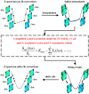
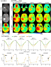
Amide proton transfer (APT)-weighted MRI is a promising molecular imaging technique that has been employed in clinic for detection and grading of brain tumors. MTRasym, the quantification method of APT, is easily influenced by B0 inhomogeneity and causes artifacts. Current model-free interpolation methods have enabled moderate B0 correction for middle offsets, but have performed poorly at limbic offsets. To address this shortcoming, we proposed a practical B0 correction approach that is suitable under time-limited sparse acquisition scenarios and for B1 ≥ 1 μT under 3T. In this study, this approach employed a simplified Lorentzian model containing only two pools of symmetric water and asymmetric solutes, to describe the Z-spectral shape with wide and ‘invisible’ CEST peaks. The B0 correction was then performed on the basis of the fitted two-pool Lorentzian lines, instead of using conventional model-free interpolation. The approach was firstly evaluated on densely sampled Z-spectra data by using the spline interpolation of all acquired 16 offsets as the gold standard. When only six offsets were available for B0 correction, our method outperformed conventional methods. In particular, the errors at limbic offsets were significantly reduced (n = 8, p < 0.01). Secondly, our method was assessed on the six-offset APT data of nine brain tumor patients. Our MTRasym (3.5 ppm), using the two-pool model, displayed a similar contrast to the vendor-provided B0-orrected MTRasym (3.5 ppm). While the vendor failed in correcting B0 at 4.3 and 2.7 ppm for a large portion of voxels, our method enabled well differentiation of B0 artifacts from tumors. In conclusion, the proposed approach could alleviate analysis errors caused by B0 inhomogeneity, which is useful for facilitating the comprehensive metabolic analysis of brain tumors.
Keywords: B0 inhomogeneity correction; Lorentzian fitting; amide proton transfer (APT) MRI; brain tumors; chemical exchange saturation transfer (CEST) MRI.
Conflict of interest statement
The authors declare no conflict of interest.
Figures


Similar articles
-
Effect of offset-frequency step size and interpolation methods on chemical exchange saturation transfer MRI computation in human brain.NMR Biomed. 2021 Apr;34(4):e4468. doi: 10.1002/nbm.4468. Epub 2021 Feb 4. NMR Biomed. 2021. PMID: 33543519
-
Amide proton transfer imaging of brain tumors using a self-corrected 3D fast spin-echo dixon method: Comparison With separate B0 correction.Magn Reson Med. 2017 Jun;77(6):2272-2279. doi: 10.1002/mrm.26322. Epub 2016 Jul 6. Magn Reson Med. 2017. PMID: 27385636
-
Systematic Evaluation of Amide Proton Chemical Exchange Saturation Transfer at 3 T: Effects of Protein Concentration, pH, and Acquisition Parameters.Invest Radiol. 2016 Oct;51(10):635-46. doi: 10.1097/RLI.0000000000000292. Invest Radiol. 2016. PMID: 27272542
-
Amide Proton Transfer-Chemical Exchange Saturation Transfer Imaging of Intracranial Brain Tumors and Tumor-like Lesions: Our Experience and a Review.Diagnostics (Basel). 2023 Feb 28;13(5):914. doi: 10.3390/diagnostics13050914. Diagnostics (Basel). 2023. PMID: 36900058 Free PMC article. Review.
-
Performance of amide proton transfer imaging to differentiate true progression from therapy-related changes in gliomas and metastases.Eur Radiol. 2025 Feb;35(2):580-591. doi: 10.1007/s00330-024-11004-y. Epub 2024 Aug 12. Eur Radiol. 2025. PMID: 39134744 Free PMC article.
References
-
- Dou W., Lin C.-Y.E., Ding H., Shen Y., Dou C., Qian L., Wen B., Wu B. Chemical exchange saturation transfer magnetic resonance imaging and its main and potential applications in pre-clinical and clinical studies. Quant. Imaging Med. Surg. 2019;9:1747–1766. doi: 10.21037/qims.2019.10.03. - DOI - PMC - PubMed
Publication types
MeSH terms
Substances
LinkOut - more resources
Full Text Sources
Medical

