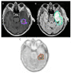
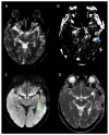
Radiotherapy Target Volume Definition in Newly Diagnosed High-Grade Glioma Using 18F-FET PET Imaging and Multiparametric MRI: An Inter Observer Agreement Study
- PMID: 36006068
- PMCID: PMC9415495
- DOI: 10.3390/tomography8040170
Radiotherapy Target Volume Definition in Newly Diagnosed High-Grade Glioma Using 18F-FET PET Imaging and Multiparametric MRI: An Inter Observer Agreement Study
Abstract
Background: The aim of this prospective monocentric study was to assess the inter-observer agreement for tumor volume delineations by multiparametric MRI and 18-F-FET-PET/CT in newly diagnosed, untreated high-grade glioma (HGG) patients. Methods: Thirty patients HGG underwent O-(2-[18F]-fluoroethyl)-l-tyrosine(18F-FET) positron emission tomography (PET), and multiparametric MRI with computation of rCBV map and K2 map. Three nuclear physicians and three radiologists with different levels of experience delineated the 18-F-FET-PET/CT and 6 MRI sequences, respectively. Spatial similarity (Dice and Jaccard: DSC and JSC) and overlap (Overlap: OV) coefficients were calculated between the readers for each sequence. Results: DSC, JSC, and OV were high for 18F-FET PET/CT, T1-GD, and T2-FLAIR (>0.67). The Spearman correlation coefficient between readers was ≥0.6 for these sequences. Cross-comparison of similarity and overlap parameters showed significant differences for DSC and JSC between 18F-FET PET/CT and T2-FLAIR and for JSC between 18F-FET PET/CT and T1-GD with higher values for 18F-FET PET/CT. No significant difference was found between T1-GD and T2-FLAIR. rCBV, K2, b1000, and ADC showed correlation coefficients between readers <0.6. Conclusion: The interobserver agreements for tumor volume delineations were high for 18-F-FET-PET/CT, T1-GD, and T2-FLAIR. The DWI (b1000, ADC), rCBV, and K2-based sequences, as performed, did not seem sufficiently reproducible to be used in daily practice.
Trial registration: ClinicalTrials.gov NCT03370926.
Keywords: 18-F-FET-PET/CT; high-grade glioma; inter-observer agreement study; multiparametric MRI; tumor volume delineation.
Conflict of interest statement
The authors of this manuscript declare no relevant conflict of interest.
Figures


Similar articles
-
Hotspot on 18F-FET PET/CT to Predict Aggressive Tumor Areas for Radiotherapy Dose Escalation Guiding in High-Grade Glioma.Cancers (Basel). 2022 Dec 23;15(1):98. doi: 10.3390/cancers15010098. Cancers (Basel). 2022. PMID: 36612093 Free PMC article.
-
Correlation between rCBV Delineation Similarity and Overall Survival in a Prospective Cohort of High-Grade Gliomas Patients: The Hidden Value of Multimodal MRI?Biomedicines. 2024 Apr 3;12(4):789. doi: 10.3390/biomedicines12040789. Biomedicines. 2024. PMID: 38672146 Free PMC article.
-
Radiotherapy target volume definition in newly diagnosed high grade glioma using 18F-FET PET imaging and multiparametric perfusion MRI: A prospective study (IMAGG).Radiother Oncol. 2020 Sep;150:164-171. doi: 10.1016/j.radonc.2020.06.025. Epub 2020 Jun 21. Radiother Oncol. 2020. PMID: 32580001
-
Comparison Between 18F-Dopa and 18F-Fet PET/CT in Patients with Suspicious Recurrent High Grade Glioma: A Literature Review and Our Experience.Curr Radiopharm. 2019;12(3):220-228. doi: 10.2174/1874471012666190115124536. Curr Radiopharm. 2019. PMID: 30644351 Review.
-
[Capabilities of 18F-FET PET/CT in a patient with brain glioma (a case report and literature review)].Zh Vopr Neirokhir Im N N Burdenko. 2018;82(2):95-99. doi: 10.17116/oftalma201882295-99. Zh Vopr Neirokhir Im N N Burdenko. 2018. PMID: 29795092 Review. Russian.
Cited by
-
Hotspot on 18F-FET PET/CT to Predict Aggressive Tumor Areas for Radiotherapy Dose Escalation Guiding in High-Grade Glioma.Cancers (Basel). 2022 Dec 23;15(1):98. doi: 10.3390/cancers15010098. Cancers (Basel). 2022. PMID: 36612093 Free PMC article.
-
Correlation between rCBV Delineation Similarity and Overall Survival in a Prospective Cohort of High-Grade Gliomas Patients: The Hidden Value of Multimodal MRI?Biomedicines. 2024 Apr 3;12(4):789. doi: 10.3390/biomedicines12040789. Biomedicines. 2024. PMID: 38672146 Free PMC article.
-
[18]F-fluoroethyl-l-tyrosine positron emission tomography for radiotherapy target delineation: Results from a Radiation Oncology credentialing program.Phys Imaging Radiat Oncol. 2024 Mar 13;30:100568. doi: 10.1016/j.phro.2024.100568. eCollection 2024 Apr. Phys Imaging Radiat Oncol. 2024. PMID: 38585372 Free PMC article.
-
Delineation and agreement of FET PET biological volumes in glioblastoma: results of the nuclear medicine credentialing program from the prospective, multi-centre trial evaluating FET PET In Glioblastoma (FIG) study-TROG 18.06.Eur J Nucl Med Mol Imaging. 2023 Nov;50(13):3970-3981. doi: 10.1007/s00259-023-06371-5. Epub 2023 Aug 11. Eur J Nucl Med Mol Imaging. 2023. PMID: 37563351 Free PMC article.
References
-
- Ostrom Q.T., Gittleman H., Liao P., Rouse C., Chen Y., Dowling J., Wolinsky Y., Kruchko C., Barnholtz-Sloan J. CBTRUS Statistical Report: Primary Brain and Central Nervous System Tumors Diagnosed in the United States in 2007–2011. Neuro-Oncology. 2014;16:iv1–iv63. doi: 10.1093/neuonc/nou223. - DOI - PMC - PubMed
-
- Law I., Albert N.L., Arbizu J., Boellaard R., Drzezga A., Galldiks N., la Fougère C., Langen K.-J., Lopci E., Lowe V., et al. Joint EANM/EANO/RANO Practice Guidelines/SNMMI Procedure Standards for Imaging of Gliomas Using PET with Radiolabelled Amino Acids and [18F]FDG: Version 1.0. Eur. J. Nucl. Med. Mol. Imaging. 2019;46:540–557. doi: 10.1007/s00259-018-4207-9. - DOI - PMC - PubMed
-
- Langen K.-J., Stoffels G., Filss C., Heinzel A., Stegmayr C., Lohmann P., Willuweit A., Neumaier B., Mottaghy F.M., Galldiks N. Imaging of Amino Acid Transport in Brain Tumours: Positron Emission Tomography with O-(2-[18F]Fluoroethyl)- L -Tyrosine (FET) Methods. 2017;130:124–134. doi: 10.1016/j.ymeth.2017.05.019. - DOI - PubMed
MeSH terms
Substances
Associated data
LinkOut - more resources
Full Text Sources
Medical

