

Bisphenol S Impairs Oestradiol Secretion during In Vitro Basal Folliculogenesis in a Mono-Ovulatory Species Model
- PMID: 36006116
- PMCID: PMC9412475
- DOI: 10.3390/toxics10080437
Bisphenol S Impairs Oestradiol Secretion during In Vitro Basal Folliculogenesis in a Mono-Ovulatory Species Model
Abstract
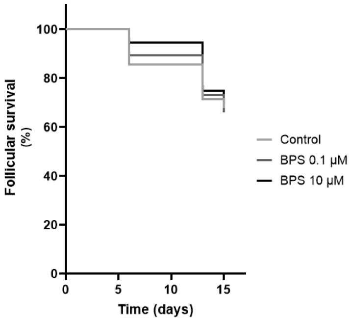
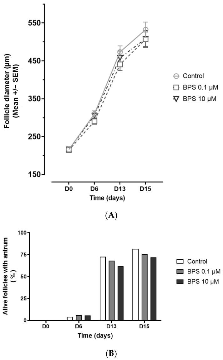
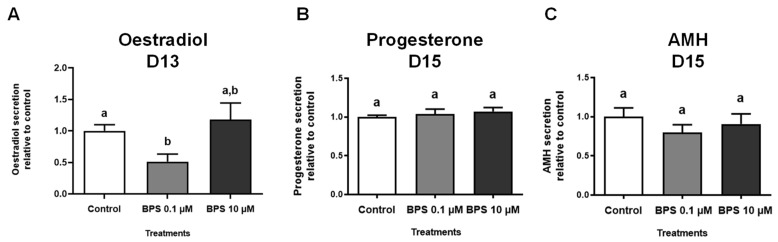
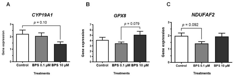
Bisphenol S (BPS) affects terminal folliculogenesis by impairing steroidogenesis in granulosa cells from different species. Nevertheless, limited data are available on its effects during basal folliculogenesis. In this study, we evaluate in vitro the effects of a long-term BPS exposure on a model of basal follicular development in a mono-ovulatory species. We cultured ovine preantral follicles (180−240 μm, n = 168) with BPS (0.1 μM (possible human exposure dose) or 10 μM (high dose)) and monitored antrum appearance and follicular survival and growth for 15 days. We measured hormonal secretions (oestradiol (at day 13 [D13]), progesterone and anti-Müllerian hormone [D15]) and expression of key follicular development and redox status genes (D15) in medium and whole follicles, respectively. BPS (0.1 µM) decreased oestradiol secretion compared with the control (−48.8%, p < 0.001), without significantly impairing antrum appearance, follicular survival and growth, anti-Müllerian hormone and progesterone secretion and target gene expression. Thus, BPS could also impair oestradiol secretion during basal folliculogenesis as it is the case during terminal folliculogenesis. It questions the use of BPS as a safe BPA substitute in the human environment. More studies are required to elucidate mechanisms of action of BPS and its effects throughout basal follicular development.
Keywords: bisphenols; endocrine disruptors; ewe; follicular growth; gene expression; hormonal secretions; ovary; plasticiser.
Conflict of interest statement
The authors declare that there are no conflict of interest that could be perceived as influencing the representation or interpretation of the reported research study.
Figures
Similar articles
-
Bisphenol A and bisphenol S both disrupt ovine granulosa cell steroidogenesis but through different molecular pathways.J Ovarian Res. 2023 Feb 3;16(1):30. doi: 10.1186/s13048-023-01114-4. J Ovarian Res. 2023. PMID: 36737804 Free PMC article.
-
Bisphenol A and S impaired ovine granulosa cell steroidogenesis.Reproduction. 2020 May;159(5):571-583. doi: 10.1530/REP-19-0575. Reproduction. 2020. PMID: 32092037
-
Bisphenol S Impaired Human Granulosa Cell Steroidogenesis in Vitro.Int J Mol Sci. 2020 Mar 6;21(5):1821. doi: 10.3390/ijms21051821. Int J Mol Sci. 2020. PMID: 32155818 Free PMC article.
-
A new chapter in the bisphenol A story: bisphenol S and bisphenol F are not safe alternatives to this compound.Fertil Steril. 2015 Jan;103(1):11-21. doi: 10.1016/j.fertnstert.2014.11.005. Epub 2014 Dec 2. Fertil Steril. 2015. PMID: 25475787 Review.
-
Endocrinologic control of normal canine ovarian function.Reprod Domest Anim. 2009 Jul;44 Suppl 2:3-15. doi: 10.1111/j.1439-0531.2009.01414.x. Reprod Domest Anim. 2009. PMID: 19754529 Review.
Cited by
-
Invisible Hand behind Female Reproductive Disorders: Bisphenols, Recent Evidence and Future Perspectives.Toxics. 2023 Dec 7;11(12):1000. doi: 10.3390/toxics11121000. Toxics. 2023. PMID: 38133401 Free PMC article. Review.
-
The Ovary as a Target Organ for New Generation Bisphenols Toxicity.Toxics. 2025 Feb 26;13(3):164. doi: 10.3390/toxics13030164. Toxics. 2025. PMID: 40137491 Free PMC article. Review.
-
Different types of bisphenols alter ovarian steroidogenesis: Special attention to BPA.Heliyon. 2023 Jun 1;9(6):e16848. doi: 10.1016/j.heliyon.2023.e16848. eCollection 2023 Jun. Heliyon. 2023. PMID: 37303564 Free PMC article.
-
Impact of Bisphenol A and its alternatives on oocyte health: a scoping review.Hum Reprod Update. 2024 Dec 1;30(6):653-691. doi: 10.1093/humupd/dmae025. Hum Reprod Update. 2024. PMID: 39277428 Free PMC article.
References
-
- Monniaux D., Cadoret V., Clément F., Dalbies-Tran R., Elis S., Fabre S., Maillard V., Monget P., Uzbekova S. Ilpo Huhtaniemi and Luciano Martini, Encyclopedia of Endocrine Diseases. 2nd ed. Volume 2. Academic Press (Elsevier); Oxford, UK: 2019. Folliculogenesis; pp. 377–398.
Grants and funding
LinkOut - more resources
Full Text Sources
