. 2022 Aug 19;14(8):566.
doi: 10.3390/toxins14080566.

Isolation, Characterization and Biological Action of Type-1 Ribosome-Inactivating Proteins from Tissues of Salsola soda L

Affiliations

Isolation, Characterization and Biological Action of Type-1 Ribosome-Inactivating Proteins from Tissues of Salsola soda L

Nicola Landi et al. Toxins (Basel). .

Abstract

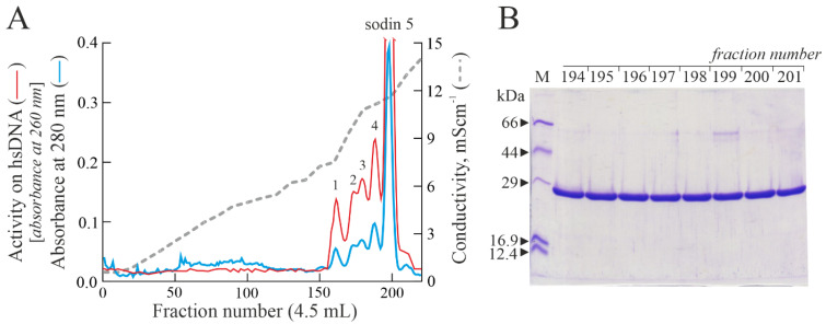
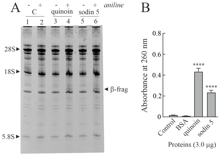
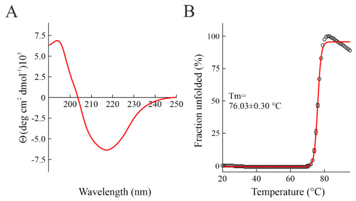
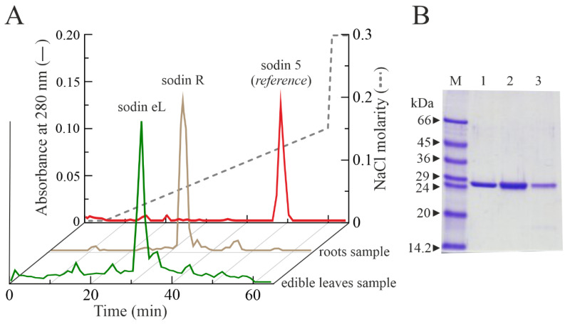
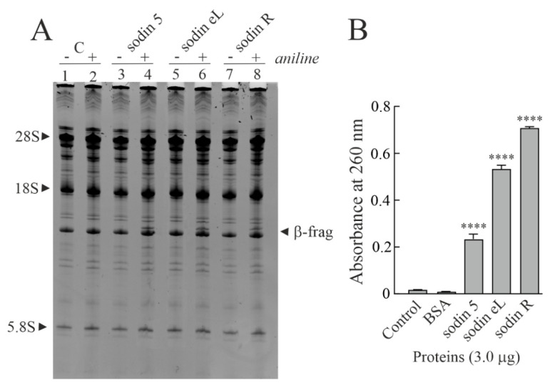
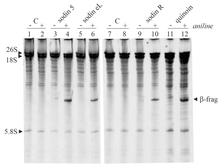
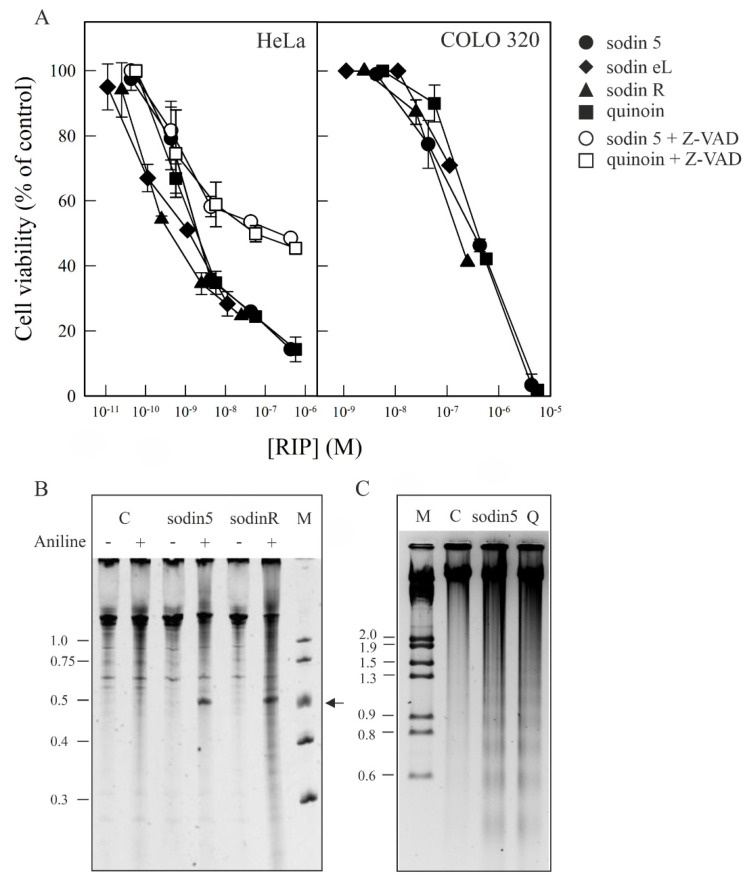
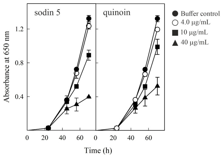
Ribosome-inactivating proteins (RIPs) are known as RNA N-glycosylases. They depurinate the major rRNA, damaging ribosomes and inhibiting protein synthesis. Here, new single-chain (type-1) RIPs named sodins were isolated from the seeds (five proteins), edible leaves (one protein) and roots (one protein) of Salsola soda L. Sodins are able to release Endo's fragment when incubated with rabbit and yeast ribosomes and inhibit protein synthesis in cell-free systems (IC50 = 4.83-79.31 pM). In addition, sodin 5, the major form isolated from seeds, as well as sodin eL and sodin R, isolated from edible leaves and roots, respectively, display polynucleotide:adenosine glycosylase activity and are cytotoxic towards the Hela and COLO 320 cell lines (IC50 = 0.41-1200 nM), inducing apoptosis. The further characterization of sodin 5 reveals that this enzyme shows a secondary structure similar to other type-1 RIPs and a higher melting temperature (Tm = 76.03 ± 0.30 °C) and is non-glycosylated, as other sodins are. Finally, we proved that sodin 5 possesses antifungal activity against Penicillium digitatum.

Keywords: agretti; antifungal activity; cytotoxicity; edible plants; protein purification; rRNA N-glycosylases.


Conflict of interest statement

The authors have no conflict of interest to declare.

Figures

Figure 1
Figure 1
(A) Elution profile after cation exchange chromatography on the CM-Sepharose column, showing five peaks (peaks 1–4 and sodin 5) with PNAG activity (arbitrary units). (B) SDS-PAGE analysis of 194–201 fractions (5.0 μg) from sodin 5 obtained after cation exchange chromatography (A). M, molecular weight markers. SDS-PAGE in the presence of β-mercaptoethanol was carried out in 12% polyacrylamide separating gel and then stained with Coomassie brilliant blue.
Figure 2
Figure 2
(A) rRNA N-glycosylase activity on rabbit ribosomes. Quinoin from C. quinoa seeds (3.0 µg; lanes 3 and 4) as a positive control and sodin 5 (3.0 µg; lanes 5 and 6) were incubated with ribosomes. Then, rRNA was extracted, treated with acid aniline and separated as described in the Materials and Methods section. (+) and (−) indicate with and without aniline treatment. ‘β-frag’ indicates the position of Endo’s fragment released by the aniline treatment of rRNA from rabbit ribosomes. (B) Polynucleotide:adenosine glycosylase activity of BSA (negative control) or quinoin and sodin 5 type-1 RIPs. Proteins (3.0 µg) were assayed on salmon sperm DNA as described in the Materials and Methods section. The mean results ± SD of three experiments performed in triplicate are reported. The data were compared to the control and analyzed by one-way ANOVA with Dunnett’s post hoc test (****, p < 0.0001).
Figure 3
Figure 3
(A) Far-UV CD spectrum of sodin 5. (B) Thermal denaturation profile of sodin 5 (concentration: 0.15 mg mL1). The fraction unfolded at 278 nm is plotted as a function of temperature. The red line represents fit curve.
Figure 4
Figure 4
(A) Elution profile after FPLC on an AKTA Purifier System from cation exchange chromatography using a Source 15S PE 4.6/100 column, showing a single protein peak from S. soda roots and edible leaves, named sodin R and sodin eL, respectively. The elution profile of sodin 5 from S. soda seeds was reported as a reference chromatographic profile. (B) SDS-PAGE analysis of fractions (5.0 μg) from sodin 5, sodin R and sodin eL (lanes 1, 2 and 3, respectively), obtained after Source 15S chromatography. M, molecular weight markers. SDS-PAGE in the presence of β-mercaptoethanol was carried out in 12% polyacrylamide separating gel and then stained with Coomassie brilliant blue.
Figure 5
Figure 5
(A) rRNA N-glycosylase activity assayed on rabbit ribosomes. Sodin 5 (3.0 μg; lanes 3 and 4) as a positive control and sodin eL (3.0 μg; lanes 5 and 6) or sodin R (3.0 μg; lanes 7 and 8) were incubated with ribosomes. Then, rRNA was extracted, treated with acid aniline and separated as described in the Materials and Methods section. (+) and (−) indicate with and without aniline treatment. ‘β-frag’ indicates the position of Endo’s fragment released by the aniline treatment of rRNA from rabbit ribosomes. (B) Polynucleotide:adenosine glycosylase activity of BSA (negative control) or sodin 5, sodin eL and sodin R type-1 RIPs. Proteins (3.0 μg) were assayed on salmon sperm DNA as described in the Materials and Methods section. The mean results ± SD of three experiments performed in triplicate are reported. Data were compared to the control and analyzed by one-way ANOVA with Dunnett’s post hoc test (****, p < 0.0001).
Figure 6
Figure 6
rRNA N-glycosylase activity assayed on yeast ribosomes. rRNA N-glycosylase activity was analyzed as reported in the Materials and Methods section. Each lane contained 5 µg of RNA isolated from either untreated (control) or RIP-treated ribosomes from yeast. (+) and (−) indicate with and without aniline treatment. ‘β-frag’ indicates the position of Endo’s fragment released by the aniline treatment of rRNA from yeast ribosomes.
Figure 7
Figure 7
Induction of cytotoxicity and apoptosis on HeLa and COLO 320 cells by sodins and quinoin (A). Effect of sodins or quinoin on the viability of HeLa (left panel) and COLO 320 (right panel) cells. Cells were grown in RPMI 1640 medium and incubated with different type-1 RIP concentrations for 48 h (HeLa) and 72 h (COLO 320), and cell viability was evaluated by a colorimetric assay, as indicated in Section 4.6 of the Materials and Methods section. To investigate the effect of Z-VAD on the viability of HeLa cells, the cells were preincubated for 3 h with Z-VAD and then incubated with different concentrations of sodin 5 or quinoin for 48 h, and cell viability was evaluated. Data represent the mean ± SD of two experiments performed in duplicate. (B) rRNA N-glycosylase activity of sodin 5 and sodin R on RNA from HeLa cells. rRNA N-glycosylase activity was evaluated as reported in the Materials and Methods section. Each lane contained 2.0 μg of RNA isolated from either untreated cells (C, control) or cells incubated with 8 nM of sodin 5 or 5 nM of sodin R for 48 h. The arrow indicates the RNA fragment released as a result of RIP action upon the acid aniline treatment. Numbers indicate the size of the standards (M) in nucleotides. (C) Effect of sodin 5 and quinoin on internucleosomal DNA fragmentation. COLO 320 cells were incubated in the absence (C, control) or presence of 0.4 μM of sodin 5 or 0.6 μM of quinoin (Q) for 72 h. The DNA was isolated, and 4.0 μg was electrophoresed, as indicated in Section 4.7. The numbers indicate the corresponding size of the standards (M) (λDNA HindIII/EcoRI) in Kb. (+) and (−) indicate with and without aniline treatment.
Figure 8
Figure 8
Antifungal activity of sodin 5 (left panel) and quinoin (right panel) against Penicillium digitatum, measured in a microtiter plate bioassay. Conidia of P. digitatum were grown in Potato Dextrose Broth (PDB) for 24 h before exposure to different RIP concentrations. Fungal growth was followed for 70 h and measured as an increase in absorbance at 650 nm. The curves represent the buffer control or different amounts (µg/mL) of both toxins. The mean results ± SE of two experiments performed in triplicate are reported.

References

    1. Endo Y., Huber P.W., Wool I.G. The ribonuclease activity of the cytotoxin alpha-sarcin. The characteristics of the enzymatic activity of alpha-sarcin with ribosomes and ribonucleic acids as substrates. J. Biol. Chem. 1983;258:2662–2667. doi: 10.1016/S0021-9258(18)32977-6. - DOI - PubMed
    1. Shi X., Khade P.K., Sanbonmatsu K.Y., Joseph S. Functional role of the sarcin-ricin loop of the 23S rRNA in the elongation cycle of protein synthesis. J. Mol. Biol. 2012;419:125–138. doi: 10.1016/j.jmb.2012.03.016. - DOI - PMC - PubMed
    1. Barbieri L., Valbonesi P., Bonora E., Gorini P., Bolognesi A., Stirpe F. Polynucleotide:adenosine glycosidase activity of ribosome-inactivating proteins: Effect on DNA, RNA and poly(A) Nucleic Acids Res. 1997;25:518–522. doi: 10.1093/nar/25.3.518. - DOI - PMC - PubMed
    1. Barbieri L., Valbonesi P., Righi F., Zuccheri G., Monti F., Gorini P., Samorí B., Stirpe F. Polynucleotide:Adenosine glycosidase is the sole activity of ribosome-inactivating proteins on DNA. J. Biochem. 2000;128:883–889. doi: 10.1093/oxfordjournals.jbchem.a022827. - DOI - PubMed
    1. Barbieri L., Gorini P., Valbonesi P., Castiglioni P., Stirpe F. Unexpected activity of saporins. Nature. 1994;372:624. doi: 10.1038/372624a0. - DOI - PubMed

Publication types

Substances

LinkOut - more resources