. 2022 Aug 1;7(8):163.
doi: 10.3390/tropicalmed7080163.

Involvement of Pore Formation and Osmotic Lysis in the Rapid Killing of Gamma Interferon-Pretreated C166 Endothelial Cells by Rickettsia prowazekii

Affiliations

Involvement of Pore Formation and Osmotic Lysis in the Rapid Killing of Gamma Interferon-Pretreated C166 Endothelial Cells by Rickettsia prowazekii

Jenifer Turco. Trop Med Infect Dis. .

Abstract

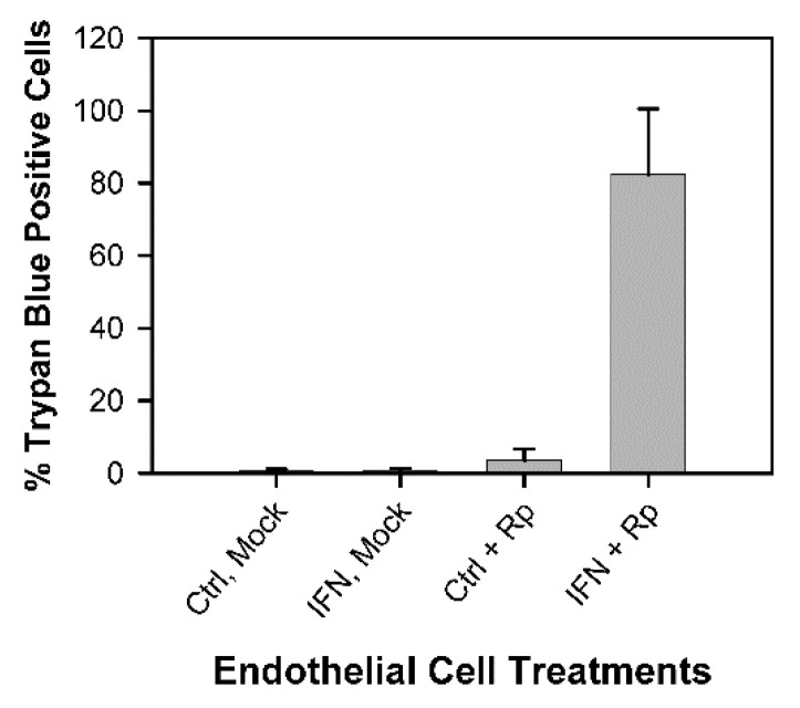
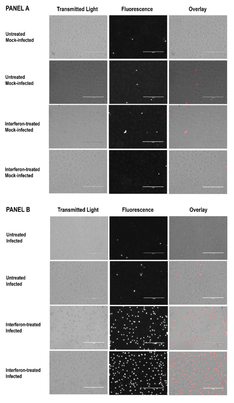
Rickettsia prowazekii, the bacterial cause of epidemic typhus in humans, proliferates mainly within the microvascular endothelial cells. Previous studies have shown that murine macrophage-like RAW264.7 cells are rapidly damaged if they are pretreated with gamma interferon (IFN-γ) and then infected with R. prowazekii. In the present study, the effects of IFN-γ and R. prowazekii on murine C166 endothelial cells were evaluated. In the IFN-γ-pretreated R. prowazekii-infected endothelial cell cultures, evidence of cell damage was observed within several hours after addition of the rickettsiae. Considerable numbers of the cells became permeable to trypan blue dye and ethidium bromide, and substantial amounts of lactate dehydrogenase (LDH) were released from the cells. Such evidence of cellular injury was not observed in the untreated infected cultures or in any of the mock-infected cultures. Polyethylene glycols (PEGs) of different nominal average molecular weights were used to assess the possible involvement of pore formation and osmotic lysis in this cellular injury. PEG 8000 dramatically suppressed LDH release, PEG 4000 partially inhibited it, and PEGs 2000 and 1450 had no effect. Despite its inhibition of LDH release, PEG 8000 did not prevent the staining of the IFN-γ-pretreated infected endothelial cells by ethidium bromide. These findings suggest that the observed cellular injury involves the formation of pores in the endothelial cell membranes, followed by osmotic lysis of the cells.

Keywords: Rickettsia prowazekii; cell death; cytokine; endothelial cell; epidemic typhus; host response; interferon; rickettsia.


Conflict of interest statement

The author declares no conflict of interest. The funders had no role in the design of the study; in the collection, analyses, or interpretation of data; in the writing of the manuscript; or in the decision to publish the results.

Figures

Figure 1
Figure 1
IFN-γ-pretreated cultures of C166 endothelial cells showed increased permeability to trypan blue dye within 4 h after addition of R. prowazekii. Data were collected in twelve independent experiments. Each bar represents the mean ± standard deviation (SD) for the following numbers of determinations: untreated mock-infected cultures (Ctrl, Mock), n = 19; IFN-γ-pretreated mock-infected cultures (IFN, Mock), n = 21; untreated R. prowazekii-infected cultures (Ctrl + Rp), n = 24; IFN-γ-pretreated R. prowazekii-infected cultures (IFN + Rp), n = 26. The IFN-γ-pretreated infected cultures differed significantly from the other cultures (p < 0.01). At 1 h after addition of the rickettsiae, the percentages of cells infected were 94.9 ± 6.1 and 96.4 ± 6.3, and the numbers of rickettsiae per infected cell were 13.6 ± 4.8 and 15.9 ± 6.4 in the untreated and IFN-γ-pretreated cultures, respectively (mean ± SD, n = 16) (Table S2).
Figure 2
Figure 2
Results of ethidium bromide staining of untreated and IFN-γ-pretreated endothelial cells 3 to 6 h after the start of the mock infection (Panel A) or after addition of R. prowazekii (Panel B). The micrographs show duplicate cultures from a representative experiment. Figure S1 shows all images from the experiment, and indicates which images are shown here. Each bar represents 200 µm. To eliminate concern about image manipulation, all transmitted light images were captured under identical conditions, and all fluorescence images were captured under identical conditions as well. All transmitted light micrographs and all overlays were then adjusted for exposure and contrast using identical settings in Adobe Photoshop CC 2018® software.
Figure 3
Figure 3
Release of lactate dehydrogenase (LDH) by IFN-γ-pretreated C166 endothelial cells after addition of R. prowazekii (Rp). Each value represents the mean ± standard deviation; the number of determinations for each point is shown in Table S3. Individual data points are shown in Table S4. At one hour after addition of the rickettsiae, the rickettsial infections were determined by microscopic counting of stained cells from triplicate cultures for each dilution of the rickettsial preparation used (1/900, RpLow; 1/450, RpMedium; and 1/200, RpHigh). The percentages of cells infected (means ± standard deviations) were 64.0 ± 9.5, 84.3 ± 5.9, and 94.3 ± 5.5, respectively, in the untreated cultures and 78.0 ± 4.4, 93.3 ± 3.2, and 96.0 ± 1.0, respectively, in the IFN-γ-pretreated cultures. The numbers of rickettsiae per infected cell were 3.7 ± 0.7, 6.9 ± 0.9, and 13.7 ± 3.7, respectively, in the untreated cultures and 6.0 ± 1.3, 9.2 ± 2.5, and 15.1 ± 1.5, respectively, in the IFN-γ-pretreated cultures (Table S5). In the panel below the figure, “Control” indicates the untreated cultures and “IFN” indicates the IFN-γ-pretreated cultures. In certain instances, the values for the standard deviations were within the symbols.
Figure 4
Figure 4
Effect of PEGs of different nominal average molecular weights on the release of lactate dehydrogenase (LDH) from IFN-γ-pretreated C166 endothelial cells after addition of R. prowazekii. PEGs were added to IFN-γ-pretreated and washed cells along with rickettsiae and samples of the culture media were collected for assay of LDH activity at various times. Table 1 provides the number of determinations and shows additional data from these experiments. Table S4 provides the individual data points. For PEG 8000, the values for the standard deviations were within the symbols. PEGs were used at a concentration of 30 mM, except for PEG 8000, which was used at a concentration of 15 mM. At 2 h, 3 h, and 4 h after addition of the rickettsiae, the mean value for the cultures without PEG differed significantly from the mean values for the cultures with PEG 8000 or PEG 4000 (p < 0.05).
Figure 5
Figure 5
Results of ethidium bromide staining of untreated and IFN-γ-pretreated endothelial cells 3 to 6 h after the start of the mock infection (Panels AD) or after addition of R. prowazekii (Panels EH). d/a, PEG 8000 (15 mM) was added during the mock infection or rickettsial infection and was continuously present thereafter; a15 min, PEG 8000 was added after the 15-min centrifugation; a60 min, PEG 8000 was added 60 min after the start of the mock infection or rickettsial infection. The micrographs in all panels show duplicate cultures from a representative experiment. Figure S2 shows all images from the experiment and indicates which images are shown here. Each bar represents 200 µm. To eliminate concern about image manipulation, all transmitted light images were captured under identical conditions, and all fluorescence images were captured under identical conditions as well. All transmitted light micrographs and all overlays were then adjusted for exposure and contrast using identical settings in Adobe Photoshop CC 2018® software.
Figure 5
Figure 5
Results of ethidium bromide staining of untreated and IFN-γ-pretreated endothelial cells 3 to 6 h after the start of the mock infection (Panels AD) or after addition of R. prowazekii (Panels EH). d/a, PEG 8000 (15 mM) was added during the mock infection or rickettsial infection and was continuously present thereafter; a15 min, PEG 8000 was added after the 15-min centrifugation; a60 min, PEG 8000 was added 60 min after the start of the mock infection or rickettsial infection. The micrographs in all panels show duplicate cultures from a representative experiment. Figure S2 shows all images from the experiment and indicates which images are shown here. Each bar represents 200 µm. To eliminate concern about image manipulation, all transmitted light images were captured under identical conditions, and all fluorescence images were captured under identical conditions as well. All transmitted light micrographs and all overlays were then adjusted for exposure and contrast using identical settings in Adobe Photoshop CC 2018® software.

Similar articles

References

    1. Fournier P.-E., Raoult D. Rickettsia. In: Trujillo M.E., Dedysh S., DeVos P., Hedlund B., Kämpfer P., Rainey F.A., Whitman W.B., editors. Bergey’s Manual of Systematics of Archaea and Bacteria. John Wiley & Sons, Inc.; Hoboken, NJ, USA: 2019. pp. 1–42. - DOI
    1. Blanton L.S. The rickettsioses, a practical update. Infect. Dis. Clin. N. Am. 2019;33:213–229. doi: 10.1016/j.idc.2018.10.010. - DOI - PMC - PubMed
    1. Salje J. Cells within cells: Rickettsiales and the obligate intracellular bacterial lifestyle. Nat. Rev. Microbiol. 2021;19:375–390. doi: 10.1038/s41579-020-00507-2. - DOI - PubMed
    1. McGinn J., Lamason R.L. The enigmatic biology of rickettsiae: Recent advances, open questions and outlook. Pathog. Dis. 2021;79:ftab019. doi: 10.1093/femspd/ftab019. - DOI - PMC - PubMed
    1. Voss O.H., Rahman M.S. Rickettsia-host interaction: Strategies of intracytosolic host colonization. Pathog. Dis. 2021;79:ftab015. doi: 10.1093/femspd/ftab015. - DOI - PMC - PubMed

LinkOut - more resources