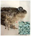
Histological Study of Glandular Variability in the Skin of the Natterjack Toad- Epidalea calamita (Laurenti, 1768)-Used in Spanish Historical Ethnoveterinary Medicine and Ethnomedicine
- PMID: 36006338
- PMCID: PMC9414601
- DOI: 10.3390/vetsci9080423
Histological Study of Glandular Variability in the Skin of the Natterjack Toad- Epidalea calamita (Laurenti, 1768)-Used in Spanish Historical Ethnoveterinary Medicine and Ethnomedicine
Abstract
Common toads have been used since ancient times for remedies and thus constitute excellent biological material for pharmacological and natural product research. According to the results of a previous analysis of the therapeutic use of amphibians in Spain, we decided to carry out a histological study that provides a complementary view of their ethnopharmacology, through the natterjack toad (Epidalea calamita). This species possesses a characteristic integument, where the parotoid glands stand out, and it has been used in different ethnoveterinary and ethnomedical practices. This histological study of their glandular variability allow us to understand the stages through which the animal synthesises and stores a heterogeneous glandular content according to the areas of the body and the functional moment of the glands. To study tegumentary cytology, a high-resolution, plastic embedding, semi-thin (1 micron) section method was applied. Up to 20 skin patches sampled from the dorsal and ventral sides were processed from the two adult specimens collected, which were roadkill. Serous/venom glands display a genetic and biochemical complexity, leading to a cocktail that remains stored (and perhaps changes over time) until extrusion, but mucous glands, working continuously to produce a surface protection layer, also produce a set of active protein (and other) substances that dissolve into mucous material, making a biologically active covering. This study provides a better understanding of the use of traditional remedies in ethnoveterinary medicine.
Keywords: Epidalea calamita; amphibian drug discovery; epistemological approach; ethnoveterinary medicine; histology; skin; zootherapy.
Conflict of interest statement
The authors declare no conflict of interest.
Figures


Similar articles
-
Differential effects of dietary protein on early life-history and morphological traits in natterjack toad (Epidalea calamita) tadpoles reared in captivity.Zoo Biol. 2013 Jul-Aug;32(4):457-62. doi: 10.1002/zoo.21067. Epub 2013 Mar 18. Zoo Biol. 2013. PMID: 23508569
-
Ethnoveterinary remedies used in the Algerian steppe: Exploring the relationship with traditional human herbal medicine.J Ethnopharmacol. 2019 Nov 15;244:112164. doi: 10.1016/j.jep.2019.112164. Epub 2019 Aug 13. J Ethnopharmacol. 2019. PMID: 31419498
-
Development of Nuclear Microsatellite Loci and Mitochondrial Single Nucleotide Polymorphisms for the Natterjack Toad, Bufo (Epidalea) calamita (Bufonidae), Using Next Generation Sequencing and Competitive Allele Specific PCR (KASPar).J Hered. 2016;107(7):660-665. doi: 10.1093/jhered/esw068. Epub 2016 Sep 15. J Hered. 2016. PMID: 27634537
-
The use of domestic animals and their derivative products in contemporary Spanish ethnoveterinary medicine.J Ethnopharmacol. 2021 May 10;271:113900. doi: 10.1016/j.jep.2021.113900. Epub 2021 Feb 5. J Ethnopharmacol. 2021. PMID: 33549762
-
Therapeutic and prophylactic uses of invertebrates in contemporary Spanish ethnoveterinary medicine.J Ethnobiol Ethnomed. 2016 Sep 5;12(1):36. doi: 10.1186/s13002-016-0111-1. J Ethnobiol Ethnomed. 2016. PMID: 27595672 Free PMC article. Review.
References
-
- Adams F. The Genuine Works of Hippocrates. Volume 1 Sydenham Society; London, UK: 1849.
-
- Schawe C.W. Veterinary Medicine and Human Health. Bailliere, Tindall and Cassell; London, UK: 1969.
-
- Wilkenson L. Animals and Disease: An Introduction to the History of Comparative Medicine. Cambridge University Press; Cambridge, UK: 1992.
-
- Cam M.-T., editor. La Médecine Vétérinaire Antique: Sources Écrites, Archéologiques, Iconographiques. Presses Universitaires; Rennes, France: 2007.
LinkOut - more resources
Full Text Sources

