. 2022 Aug 25;79(9):496.
doi: 10.1007/s00018-022-04527-4.

Molecular landscape of BoNT/B bound to a membrane-inserted synaptotagmin/ganglioside complex

Affiliations

Molecular landscape of BoNT/B bound to a membrane-inserted synaptotagmin/ganglioside complex

Jorge Ramirez-Franco et al. Cell Mol Life Sci. .

Abstract

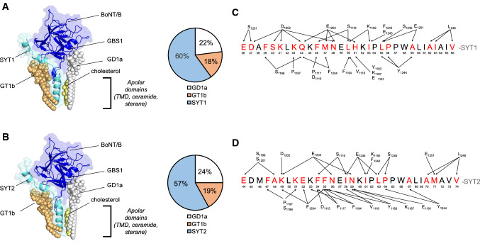
Botulinum neurotoxin serotype B (BoNT/B) uses two separate protein and polysialoglycolipid-binding pockets to interact with synaptotagmin 1/2 and gangliosides. However, an integrated model of BoNT/B bound to its neuronal receptors in a native membrane topology is still lacking. Using a panel of in silico and experimental approaches, we present here a new model for BoNT/B binding to neuronal membranes, in which the toxin binds to a preassembled synaptotagmin-ganglioside GT1b complex and a free ganglioside allowing a lipid-binding loop of BoNT/B to interact with the glycone part of the synaptotagmin-associated GT1b. Furthermore, our data provide molecular support for the decrease in BoNT/B sensitivity in Felidae that harbor the natural variant synaptotagmin2-N59Q. These results reveal multiple interactions of BoNT/B with gangliosides and support a novel paradigm in which a toxin recognizes a protein/ganglioside complex.

Keywords: Botulinum neurotoxin type B; Gangliosides; Molecular modelling; Synaptotagmin.


Conflict of interest statement

The authors declare that they have no conflict of interest.

Figures

Fig. 1
Fig. 1
SPR based on-chip reconstitution of BoNT/B binding to SYT JMD pre-assembled with GT1b. A GT1b binding to pSYT1 (Biot-GEGKEDAFSKLKQKFMNELHKIPLPPW) and pSYT2 (Biot-GESQEDMFAKLKDKFFNEINKIPLPPW). GT1b (200 nM) was injected for 1 min over immobilized pSYT1, pSYT2 and pSYT9 (Biot-HDSCQDFIYHLRDRARPRLRDPDISVS) (240 RU) at 40 µl/min. Representative of > 10 independent experiments. B GT1b bound to SYT induces an increment in BoNT/B binding signal. BoNT/B (30 nM) was injected (first arrow) onto pSYT1 (260 RU) showing a transient interaction that rapidly returns to baseline level (lower dashed line). GT1b (10 nM, second arrow) was then stably immobilized on pSYT1 (ΔGT1b) generating a new baseline (upper dashed line) and BoNT/B (30 nM, third arrow) was then injected again. Black bars highlight the BoNT/B injection phases. Representative of 6 independent experiments. C GT1b potentiation of BoNT/B binding to pSYT1 depends on the amount of GT1b bound to pSYT1. Sensorgrams resulting from the interaction of BoNT/B (30 nM) with pSYT1 (300 RU) pre-assembled with various amounts of GT1b (from 0 to 400 RU) were superposed. Representative of three independent experiments
Fig. 2
Fig. 2
Overall structure of the energy-minimized complex between BoNT/B and its membrane ligands. A Model of SYT1-BoNT/B complex (SYT1 aa 34–72, BoNT/B HC aa 1079–1290). B Model of SYT2-BoNT/B complex (SYT2 aa 42–80, BoNT/B HC aa 1079–1290). The models are based on the initial superposition of a preformed synaptotagmin1/2-GT1b complex positioned in the SYT binding site of the toxin, also bound to GD1a, with a cholesterol molecule positioned between SYT-TM and GD1a ceramide. Both BoNT/B and SYT are represented as cartoons (dark and light blue respectively). The gangliosides and cholesterol are represented as spheres (GT1b: light orange, GD1a: white, cholesterol: light yellow). The apolar domains indicated in the models correspond to the sterane and isooctyl chains of cholesterol, the ceramide part of GD1a and GT1b, and the TMD of SYT. The pie charts indicate the relative distribution of the energies of interaction in the complex between BoNT/B and SYT1/2, GT1b, GD1a and between BoNT/B and SYT2, GT1b, GD1a. Note that cholesterol does not interact with the toxin, but with SYT TM and the ceramide part of GD1a. C and D depict mapping of the intermolecular interactions between BoNT/B and SYT. C SYT1 and D SYT2 residues interacting with the toxin are highlighted in red. Arrows indicate BoNT/B-SYT interaction points. A cut off < 3.5 Å was used to select the illustrated residues
Fig. 3
Fig. 3
Close-up view of the molecular interface of SYT1-F46-M47 and SYT2-F54-F55. A Superposition of the SYT1-BoNT/B complex from PDB: 6G5K (BoNT/B in green and SYT1 in white) and the present model (BoNT/B in blue and SYT1 in light blue). B Superposition of the SYT2-BoNT/B complex from PDB 2NP0 (BoNT/B in green and SYT2 in white) and the present model (BoNT/B in blue and SYT2 in light blue). Interacting BoNT/B residues are shadowed in gray. Note that the position of BoNT/B residues are conserved between the proposed models and the corresponding crystal structures while their relative partners shift from F46 to M47 in SYT1 and F54 to F55 in SYT2.
Fig. 4
Fig. 4
Schematic overview of intermolecular interactions between GT1b-BoNT/B and GT1b-SYT. The amino acids of SYT1 (A) and SYT2 (B) are boxed while those of the toxin are circled. A cut off < 3.5 Å was used to select the residues indicated in the figure. Glc glucose, Gal galactose, Gal-Nac N- acetylgalactosamine, Sia sialic acid, Cer ceramide. Only residues with energy ≥ 3 kJ/mol are listed
Fig. 5
Fig. 5
Measurement of BoNT/B apolar loop (LBL) interaction with GT1b using Langmuir monolayers. A Stable monolayers of GT1b, lyso-LacCer (Lyso-LC), and sphingomyelin (SM) were prepared at the air–water interface at an initial surface pressure of 15–20 mN m−1. After equilibrium of the monolayer, the BoNT/B apolar loop (RFYESGIVFEEYKDY) (aa 1242–1256) was added at a final concentration of 10 µM. The data show the surface pressure increase ∆π induced by the loop as a function of time. The data are representative of three distinct experiments. B Histograms comparing the endpoints in a ± SD of three independent experiments
Fig. 6
Fig. 6
Mutations in the BoNT/B binding interface of SYTs decrease the binding of BoNT/B to HEK 293 cells. A Immunostaining of SYT1 (green) and BoNT/B (red) in HEK 293 cells transfected with either SYT1-WT (top) or H51G-SYT1 (bottom). DAPI signal is shown in blue, and the merge over DIC images indicated. Orthogonal projections of SYT1 labelling (green) in cells transfected with WT-SYT1 or SYT1-H51G (right). Scale bars, 10 µm. B Immunostaining of SYT2 (green) and BoNT/B (red) in HEK 293 cells transfected with either SYT2-WT (top panels) or SYT2-N59Q (bottom panels). DAPI signal is shown in blue, and the merge over DIC images indicated. Orthogonal projections of SYT2 labelling (green) in cells transfected with SYT2-WT or SYT2-N59Q (right). Scale bars, 10 µm. C Quantification of BoNT/B binding (gray and pink) and SYT1 expression (black and red) in cells expressing SYT1-WT or SYT1-H51G. The number of ROIs analyzed is indicated within each column. Normalized immunoreactivity data (IR) are expressed as mean ± SEM. Mann–Whitney U test was used for comparisons. **P < 0.01; n.s. non-significant. SYT1-WT IR to SYT1-H51G IR P = 0.64; BoNT/B SYT1-WT to BoNT/B SYT1-H51G P = 4.42 × 10–12. D Quantification of BoNT/B binding (gray and pink) and SYT2 expression (black and red) in cells expressing SYT2-WT or SYT2-N59Q. The number of ROIs analyzed is indicated within each column. Data are expressed as mean ± SEM. Mann–Whitney U test was used for comparisons. **P < 0.01; n.s. non-significant. SYT2-WT IR to SYT2-N59Q IR P = 0.64; BoNT/B SYT2-WT to BoNT/B SYT2-N59Q P < 0.001

Similar articles

Cited by

References

    1. Poulain B, Lemichez E, Popoff MR. Neuronal selectivity of botulinum neurotoxins. Toxicon. 2020;178:20–32. doi: 10.1016/j.toxicon.2020.02.006. - DOI - PubMed
    1. Dong M, Masuyer G, Stenmark P. Botulinum and tetanus neurotoxins. Annu Rev Biochem. 2019;88:811–837. doi: 10.1146/annurev-biochem-013118-111654. - DOI - PMC - PubMed
    1. Davies JR, Liu SM, Acharya KR. Variations in the botulinum neurotoxin binding domain and the potential for novel therapeutics. Toxins (Basel) 2018 doi: 10.3390/toxins10100421. - DOI - PMC - PubMed
    1. Pellett S, Yaksh TL, Ramachandran R. Current status and future directions of botulinum neurotoxins for targeting pain processing. Toxins (Basel) 2015;7:4519–4563. doi: 10.3390/toxins7114519. - DOI - PMC - PubMed
    1. Pirazzini M, Rossetto O, Eleopra R, Montecucco C. Botulinum neurotoxins: biology, pharmacology, and toxicology. Pharmacol Rev. 2017;69:200–235. doi: 10.1124/pr.116.012658. - DOI - PMC - PubMed

Grants and funding