

Biomechanical study of anterior and posterior pelvic rings using pedicle screw fixation for Tile C1 pelvic fractures: Finite element analysis
- PMID: 36006983
- PMCID: PMC9409507
- DOI: 10.1371/journal.pone.0273351
Biomechanical study of anterior and posterior pelvic rings using pedicle screw fixation for Tile C1 pelvic fractures: Finite element analysis
Abstract
Objective: The purpose of this study was to analyse the biomechanical characteristics of pedicle screws with different placement methods and diameters in the treatment of Tile C1 pelvic fractures by finite element simulation technology and to compare them with the plate fixation model to verify the effectiveness of pedicle screw fixation.
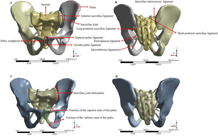
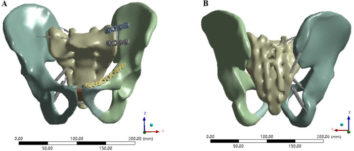
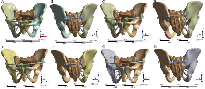
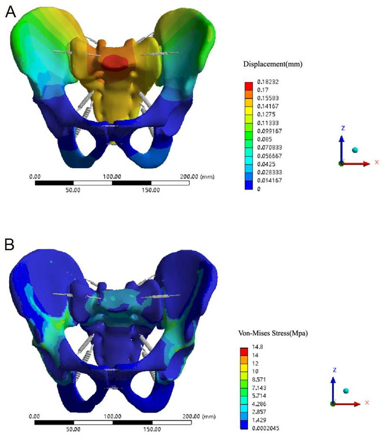
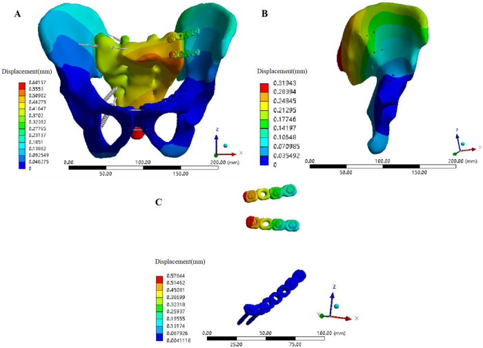
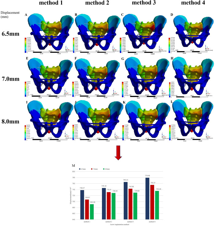
Methods: A three-dimensional digital model of a normal pelvis was obtained using computed tomography images. A finite element model of a normal pelvis containing major ligaments was built and validated (Model 1). Based on the verified normal pelvis finite element model, a Tile C1 pelvic fracture model was established (Model 2), and then a plate fixation model (Model 3) and a pedicle screw fixation model with different screw placement methods and diameters were established (Models 4-15). For all pelvic fracture fixation models, a vertical load of 500 N was applied on the upper surface of the sacrum to test the displacement and stress distribution of the pelvis in the standing state with both legs.
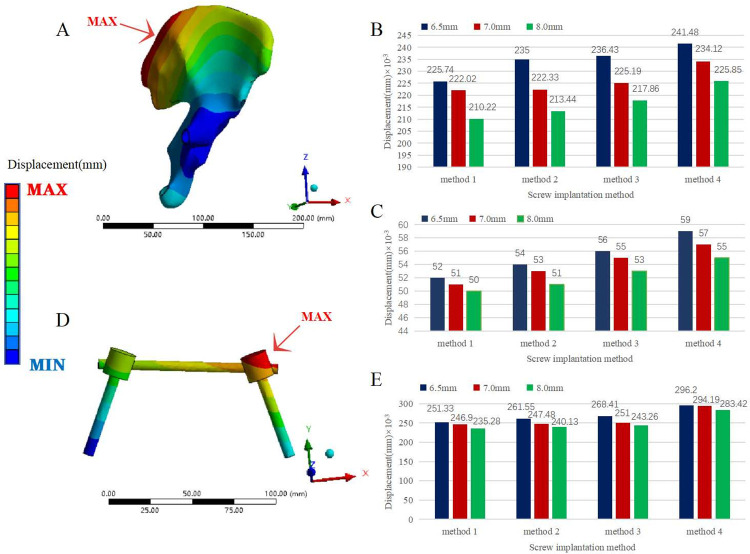
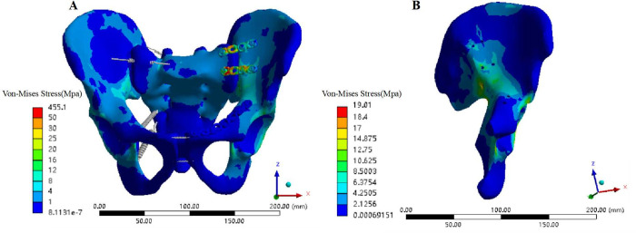
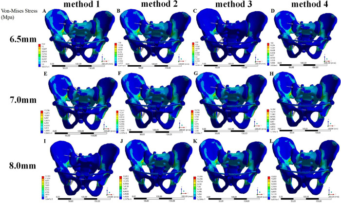
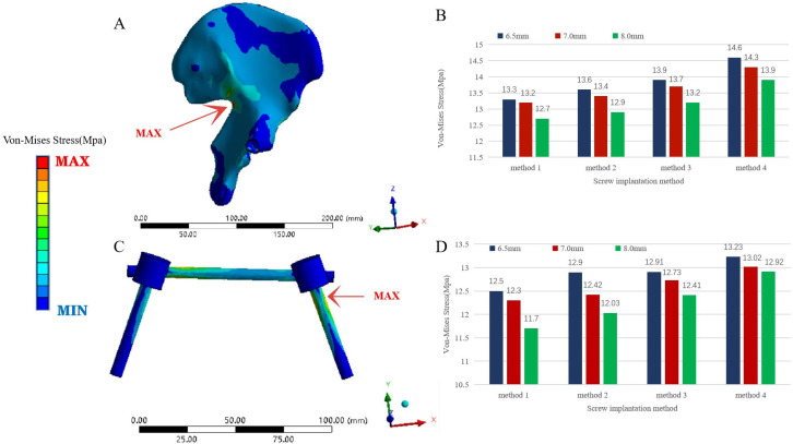
Results: The finite element simulation results showed the maximum displacement of Model 1 and Models 3-15 to be less than 1 mm. The overall maximum displacement of Models 4-15 was slightly larger than that of Model 3 (the maximum difference was 177.91×10⁻³ mm), but the maximum displacement of iliac bone and internal fixation in Models 4-15 was smaller than that of Model 3. The overall maximum stress (maximum stress of the ilium) and maximum stress of internal fixation in Models 4-15 were less than those in Model 3. The maximum displacement difference and maximum stress difference at the fracture of the pubic ramus between each fixed model were less than 0.01 mm and 1 MPa, respectively. The greater the diameter and number of pedicle screws were, the smaller the maximum displacement and stress of the pelvic fracture models were.The maximum displacement and stress of the pelvic fracture models of the screws placed on the injured side of the pubic region were smaller than the screws on the healthy side.
Conclusion: Both the anterior and posterior pelvic rings are fixed with a pedicle screw rod system for treatment of Tile C1 pelvic fractures, which can obtain sufficient biomechanical stability and can be used as a suitable alternative to other implants.The greater the diameter and number of pedicle screws were, the greater the pelvic stability was, and the greater was the stability of the screws placed on the injured side of the pubic region than the screws on the healthy side.
Conflict of interest statement
The authors have declared that no competing interests exist.
Figures
Similar articles
-
Numerical study of pedicle screw construction and locking compression plate fixation in posterior pelvic ring injuries: Analyzed by finite element method.Medicine (Baltimore). 2024 May 17;103(20):e38258. doi: 10.1097/MD.0000000000038258. Medicine (Baltimore). 2024. PMID: 38758846 Free PMC article.
-
Finite element analysis of retrograde superior ramus screw of pubis for the treament of pelvic anterior ring fracture.J Orthop Surg Res. 2025 Mar 12;20(1):263. doi: 10.1186/s13018-025-05676-5. J Orthop Surg Res. 2025. PMID: 40069735 Free PMC article.
-
Biomechanical comparative study on external fixators of new configurations in the treatment of Tile C pelvic injury.Sci Rep. 2024 Apr 25;14(1):9544. doi: 10.1038/s41598-024-60341-x. Sci Rep. 2024. PMID: 38664538 Free PMC article.
-
The Anterior Subcutaneous Pelvic Fixator (INFIX) in an Anterior Posterior Compression Type 3 Pelvic Fracture.J Orthop Trauma. 2016 Aug;30 Suppl 2:S21-2. doi: 10.1097/BOT.0000000000000583. J Orthop Trauma. 2016. PMID: 27441929 Review.
-
[Development and current situation of reconstruction methods following total sacrectomy].Zhongguo Xiu Fu Chong Jian Wai Ke Za Zhi. 2018 May 15;32(5):513-518. doi: 10.7507/1002-1892.201712054. Zhongguo Xiu Fu Chong Jian Wai Ke Za Zhi. 2018. PMID: 29806335 Free PMC article. Review. Chinese.
Cited by
-
Semi-automated finite element analyses of surgically treated acetabular fractures to investigate the biomechanical behaviour of patient-specific compared to conventional implants.J Orthop Surg Res. 2024 Sep 5;19(1):541. doi: 10.1186/s13018-024-04957-9. J Orthop Surg Res. 2024. PMID: 39237975 Free PMC article.
-
Biomechanical performance evaluation of S2AI combine with LC-2 screw for day II pelvic crescent fracture dislocation via finite element analysis.Sci Rep. 2025 May 14;15(1):16765. doi: 10.1038/s41598-025-00156-6. Sci Rep. 2025. PMID: 40368891 Free PMC article.
-
[Finite element analysis of five internal fixation modes in treatment of Day type Ⅱcrescent fracture dislocation of pelvis].Zhongguo Xiu Fu Chong Jian Wai Ke Za Zhi. 2023 Oct 15;37(10):1205-1213. doi: 10.7507/1002-1892.202306043. Zhongguo Xiu Fu Chong Jian Wai Ke Za Zhi. 2023. PMID: 37848314 Free PMC article. Chinese.
-
Modified screw-rod fixation for management of posterior pelvic ring fractures: a retrospective study.BMC Surg. 2024 Nov 20;24(1):364. doi: 10.1186/s12893-024-02654-2. BMC Surg. 2024. PMID: 39563301 Free PMC article.
-
Numerical study of pedicle screw construction and locking compression plate fixation in posterior pelvic ring injuries: Analyzed by finite element method.Medicine (Baltimore). 2024 May 17;103(20):e38258. doi: 10.1097/MD.0000000000038258. Medicine (Baltimore). 2024. PMID: 38758846 Free PMC article.
References
Publication types
MeSH terms
LinkOut - more resources
Full Text Sources
Medical
