. 2022 Sep 16;377(6612):1290-1298.
doi: 10.1126/science.abg6621. Epub 2022 Aug 25.

Lysosomal GPCR-like protein LYCHOS signals cholesterol sufficiency to mTORC1

Affiliations

Lysosomal GPCR-like protein LYCHOS signals cholesterol sufficiency to mTORC1

Hijai R Shin et al. Science. .

Abstract

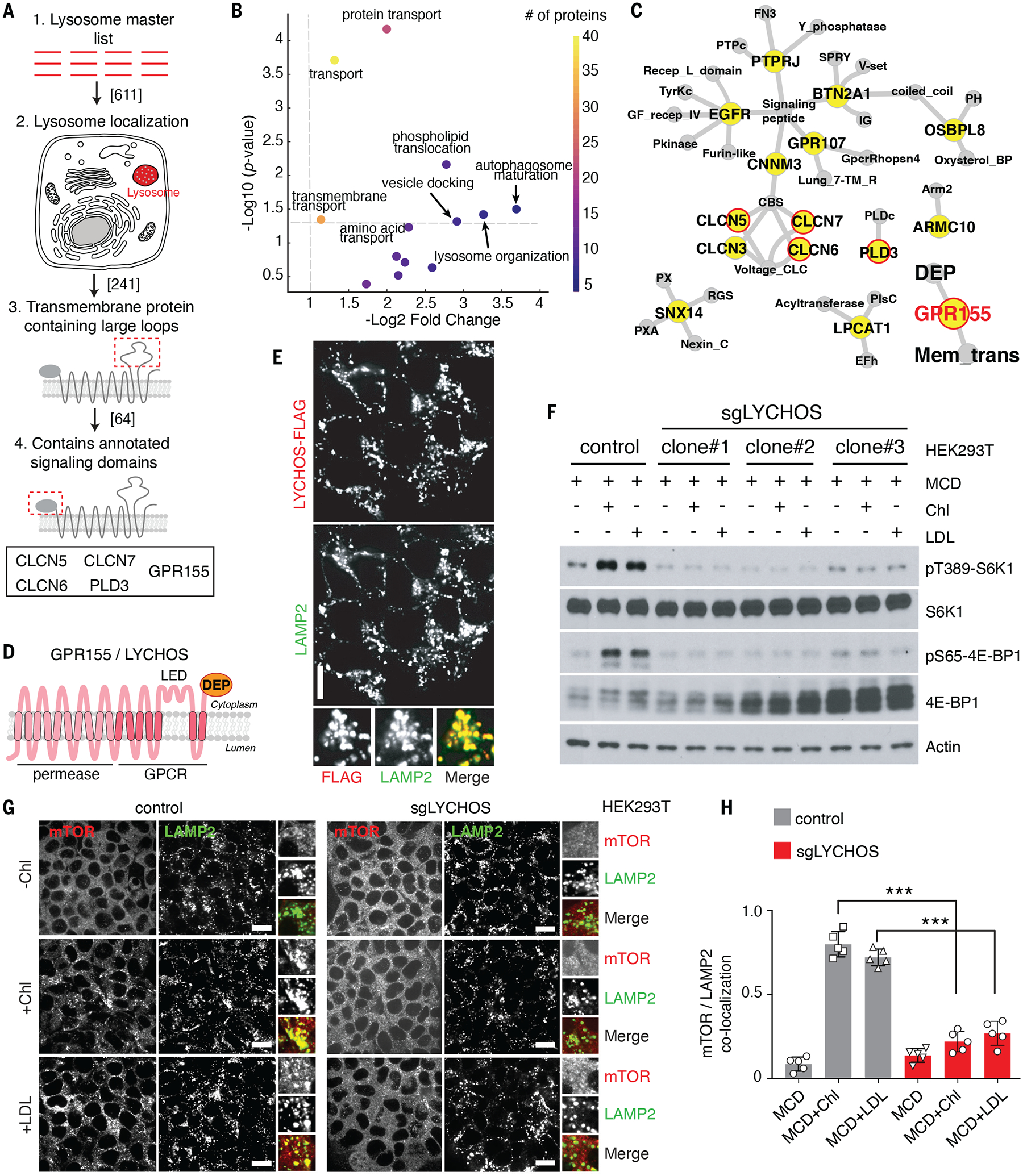
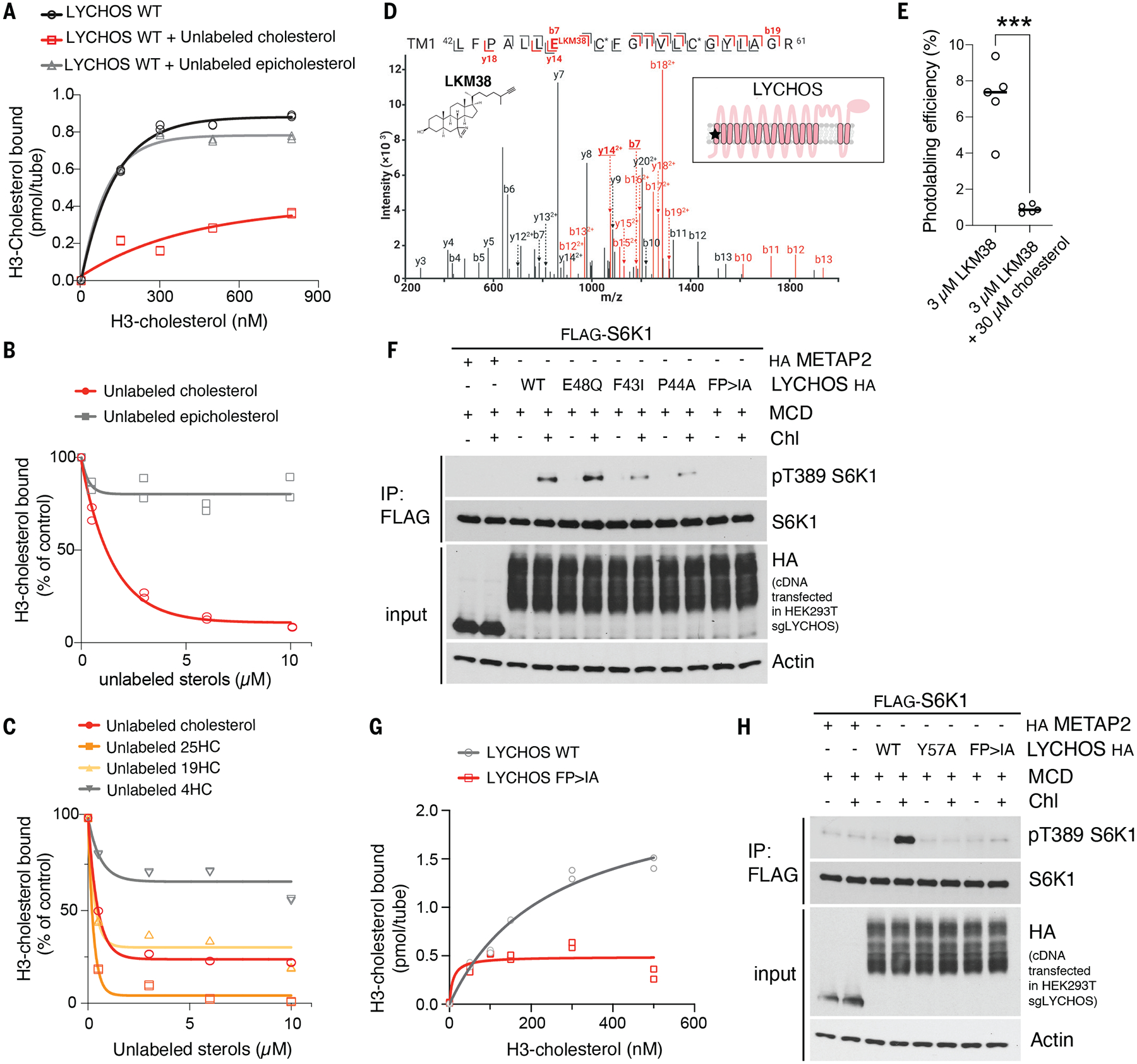
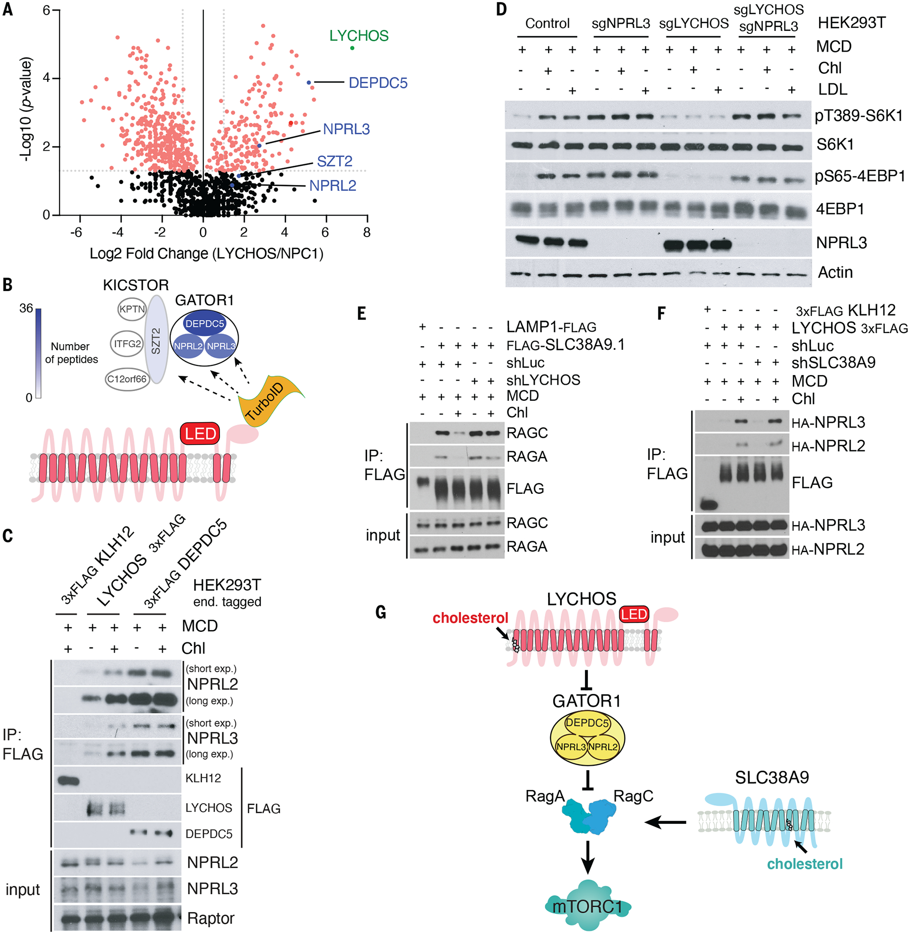
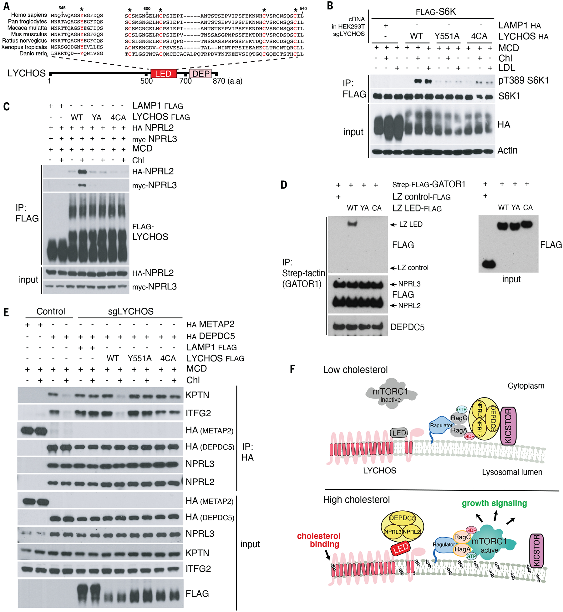
Lysosomes coordinate cellular metabolism and growth upon sensing of essential nutrients, including cholesterol. Through bioinformatic analysis of lysosomal proteomes, we identified lysosomal cholesterol signaling (LYCHOS, previously annotated as G protein-coupled receptor 155), a multidomain transmembrane protein that enables cholesterol-dependent activation of the master growth regulator, the protein kinase mechanistic target of rapamycin complex 1 (mTORC1). Cholesterol bound to the amino-terminal permease-like region of LYCHOS, and mutating this site impaired mTORC1 activation. At high cholesterol concentrations, LYCHOS bound to the GATOR1 complex, a guanosine triphosphatase (GTPase)-activating protein for the Rag GTPases, through a conserved cytoplasm-facing loop. By sequestering GATOR1, LYCHOS promotes cholesterol- and Rag-dependent recruitment of mTORC1 to lysosomes. Thus, LYCHOS functions in a lysosomal pathway for cholesterol sensing and couples cholesterol concentrations to mTORC1-dependent anabolic signaling.


Conflict of interest statement

Competing interests: R.Z. is a co-founder, stockholder, and scientific advisor for Frontier Medicines Corp., and a scientific advisor for Nine Square Therapeutics and Apertor Pharmaceuticals. D.K. N. is a co-founder, shareholder, and adviser for Frontier Medicines and Vicinitas Therapeutics. D.K.N. is also on the scientific advisory board of The Mark Foundation for Cancer Research, Photys Therapeutics, and Apertor Pharmaceuticals, and is a consultant for MPM Capital and Droia Ventures. The other authors declare that they have no competing interests.

Figures

Fig. 1.
Fig. 1.. Lysosomal transmembrane protein LYCHOS is required for cholesterol-mediated mTORC1 activation
(A) Summary chart of the workflow for the identification of lysosomal transmembrane signaling proteins.. (B) Volcano plots of ‘biological process’ GO terms enriched in lysosome-resident transmembrane proteins relative to all transmembrane proteins. (C) Network representation of lysosome-associated transmembrane proteins with large loops (blue). Gray nodes show annotated signaling domains. Predicted lysosome-resident transmembrane proteins are circled in red. (D) Schematic of the predicted GPR155/LYCHOS topology and domain organization. (E) LYCHOS is a lysosomal protein. HEK293T cells stably expressing LYCHOS-FLAG was fixed and stained with antibodies targeting FLAG and LAMP2. Scale bar, 10 μm. (F) LYCHOS is required for mTORC1 activation by cholesterol. Control HEK293T cells or LYCHOS-deleted cells (sgLYCHOS) were depleted of sterols using methyl-β-cyclodextrin (MCD, 0.75% w/v) for 2 hours, followed by re-feeding for 2 hours with 50 μM cholesterol (chl) in complex with 0.1% MCD or with 50 μg/ml LDL. Cell lysates were blotted with the indicated antibodies. (G) LYCHOS is required for cholesterol-dependent mTORC1 recruitment to lysosomes. LYCHOS-deleted HEK293T cells were subject to cholesterol depletion and restimulation, followed by immunofluorescence of endogenous mTOR and LAMP2. Representative images are shown. Scale bars, 10 μm. (H) Quantification of co-localization of mTOR with LAMP2-positive lysosomes in the indicated genotypes and conditions. Data are mean ±s.d. Statistical analysis was performed using ANOVA with Dunnett’s multiple comparison test; ***p<0.001
Fig. 2.
Fig. 2.. Cholesterol binding at the N-terminal region of LYCHOS is essential for mTORC1 activation
(A) [3H]-cholesterol binding to LYCHOS WT. 150ng of purified LYCHOS was incubated with indicated concentration of [3H]-cholesterol, in the presence or absence of 10μM cold cholesterol or epicholesterol. Bound [3H]-cholesterol was measured by scintillation counting. The assay was performed in duplicates and each data point is shown. (B,C) Competitive binding of unlabeled sterols to LYCHOS. 150ng LYCHOS was incubated with 500nM [3H]-cholesterol along with increasing concentrations of the indicated unlabeled sterol. Bound [3H]-cholesterol was measured by scintillation counting. The assay was performed in duplicates and each data point is shown. (D) CID product ion spectrum of the TM1 tryptic peptide photolabeled with 3μM LKM38. TM1 peptide (m/z=934.53, z=3) is photolabeled by LKM38 at E48. Red and black indicate product ions that do or do not contain LKM38 adduct, respectively. The C* indicates that the cysteine is alkylated by NEM. The inset schematic of GPR155 in the panel indicates the approximate location of the residues labeled by LKM38 (red star). The numerical data are included in Table S3. NEM, N-ethylmaleimide; TM, transmembrane helix. (E) Photolabeling efficiency of recombinant LYCHOS by LKM38 in the absence or presence of excess unlabeled cholesterol. Data are mean of ±s.d of n=5. Statistical analysis was performed using student t-test; ***p<0.001 (F) LYCHOS TM1 is required for cholesterol-mediated mTORC1 activation. HEK293T/sgLYCHOS were transfected with FLAG-S6K1 along with HA-tagged METAP2 (neg. control), LYCHOS WT and TM1 mutants. Cells were cholesterol-starved, or starved and restimulated as indicated in the presence of 50μM mevalonate and mevastatin, followed by FLAG immunoprecipitation (IP) and immunoblotting for the phosphorylation state and levels of the indicated proteins (G) [3H]-cholesterol binding to LYCHOS WT and LYCHOS FP>IA mutant. 150ng of purified LYCHOS were incubated with indicated concentration of [3H]-cholesterol, and bound radioactive cholesterol was measured by scintillation counting. The assay was performed in duplicates and each data point is shown. (H) LYCHOS TM1 mutants FP>IA and Y57A blunt cholesterol-mediated mTORC1 activation. HEK293T/ sgLYCHOS cells were transfected with FLAG-S6K1 along with HA-METAP2 (neg. control) or LYCHOS WT, Y57A or FP>IA-HA and analyzed as in (F).
Fig. 3.
Fig. 3.. LYCHOS promotes mTORC1 signaling via cholesterol-regulated interaction with GATOR1.
(A) TurboID-based proximity labeling combined with LC-MS/MS identified GATOR1 complex components (DEPDC5, NPRL2, and NPRL3) as interactors of LYCHOS. Volcano plot of the ratio of LYCHOS-FLAG-TurboID (LYCHOS) to NPC1-FLAG-TurboID (NPC1) is shown. Proteins with statistically significant (p value≥0.05, two-tailed unpaired t test) with fold change LYCHOS/NPC1≥2 are displayed as red circles. (B) Cartoon summarizing the TurboID proteomic analysis in (A). GATOR1 and KICSTOR subunits are color-coded according to their peptide counts. (C) Cholesterol strengthens the LYCHOS-GATOR1 interaction. HEK293T cells bearing endogenously 3xFLAG-tagged KLH12, LYCHOS and DEPDC5 were depleted of sterols then re-fed with 50 μM cholesterol, followed by FLAG immunoprecipitation and immunoblotting for the indicated proteins. (D) LYCHOS regulates cholesterol-dependent mTORC1 signaling via GATOR1. HEK293T cells lacking GATOR1 (sgNPRL3), LYCHOS (sgLYCHOS) or both (sgNPRL3/sgLYCHOS) were cholesterol-starved, or starved and refed with 50 μM cholesterol or 50 μg/ml LDL, followed by immunoblotting for the indicated proteins. (E) LYCHOS functions at upstream of RagA GTP loading. HEK293T cells stably expressing LAMP1-FLAG or FLAG-SLC38A9.1 were infected with shRNA targeting luciferase or LYCHOS for FLAG immunoprecipitation to assess SLC38A9.1-RAG A/C interaction. (F) SLC38A9.1 functions at downstream of RagA GTP loading. SLC38A9.1 was knockdown in HEK293T cells endogenously 3xFLAG-tagged KLH12 and LYCHOS cells, followed by FLAG immunoprecipitation and immunoblotting for the indicated proteins. (G)) LYCHOS and SLC38A9 mediate distinct cholesterol-sensing pathways, converging on mTORC1.
Fig. 4.
Fig. 4.. Cholesterol disrupts the GATOR1-KICSTOR interaction via the LYCHOS LED.
(A) Sequence alignment of LYCHOS LED domain. Highly conserved residues selected for mutagenesis are highlighted in red. (B) HEK293T/ sgLYCHOS cells were transfected with FLAG-S6K1 along with LAMP1-HA (neg. control) or LYCHOS WT, Y551A or 4CA-HA, followed by FLAG immunoprecipitation and immunoblotting for the phosphorylation state and levels of the indicated proteins. (C) LYCHOS LED mutations disrupt cholesterol-dependent LYCHOS interaction with GATOR1. HEK293T/sgLYCHOS cells were reconstituted with the indicated WT and LED-mutant LYCHOS constructs along with GATOR1. FLAG immunoprecipitates were analyzed by immunoblotting. (D) LYCHOS LED is sufficient for GATOR1 interaction: in vitro binding assay between purified GATOR1 and recombinant, wild-type or mutant LYCHOS LED fused to a leucine zipper (LZ). LZ alone was used as negative control. (E) In high cholesterol, LYCHOS disrupts the GATOR1-KICSTOR interaction via its LED. Control HEK293T cells, or HEK293T/sgLYCHOS cells reconstituted with the indicated WT and mutant FLAG-LYCHOS constructs, and co-expressing HA-METAP2 or HA-DEPDC5 as indicated, were cholesterol-starved, or starved and restimulated, followed by HA immunoprecipitation. Lysates were analyzed by immunoblotting (F) Molecular mechanisms of LYCHOS-dependent regulation of cholesterol-mTORC1 signaling. Under low cholesterol, stable GATOR1-KICSTOR complex promotes GATOR1-dependent GTP hydrolysis of RagA/B, which maintains mTORC1 inactive in the cytosol. In high cholesterol, a conformational change in the LED stimulates LYCHOS interaction with GATOR1 while displacing KICSTOR, thus favoring the GTP-loaded state of RagA/B that leads to lysosomal recruitment and activation of mTORC1.

Comment in

References

    1. Luo J, Yang H, Song B-L, Nat Rev Mol Cell Biol. 21, 225–245 (2020). - PubMed
    1. Riscal R, Skuli N, Simon MC, Mol Cell. 76, 220–231 (2019). - PMC - PubMed
    1. Radhakrishnan A, Goldstein JL, McDonald JG, Brown MS, Cell Metab. 8, 512–521 (2008). - PMC - PubMed
    1. Yan R et al., Science (2021), doi:10.1126/science.abb2224. - DOI
    1. Düvel K et al., Mol. Cell 39, 171–183 (2010). - PMC - PubMed

MeSH terms