Review
. 2022 Oct:90:97-109.
doi: 10.1016/j.clinimag.2022.07.005. Epub 2022 Jul 23.

Black Fungus and beyond: COVID-19 associated infections

Affiliations
Review

Black Fungus and beyond: COVID-19 associated infections

Sanaz Katal et al. Clin Imaging. 2022 Oct.

Abstract

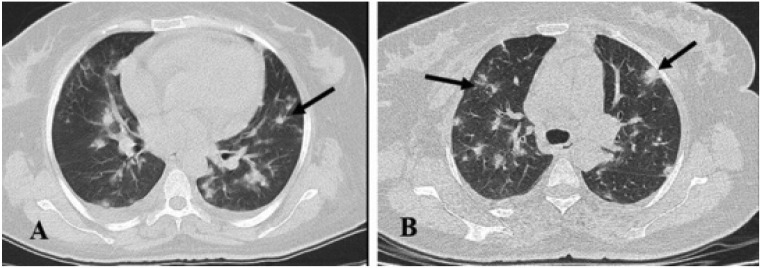
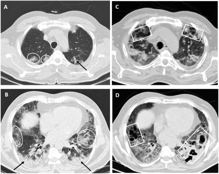
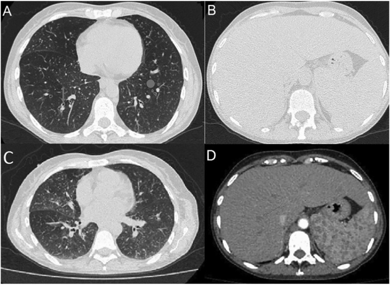
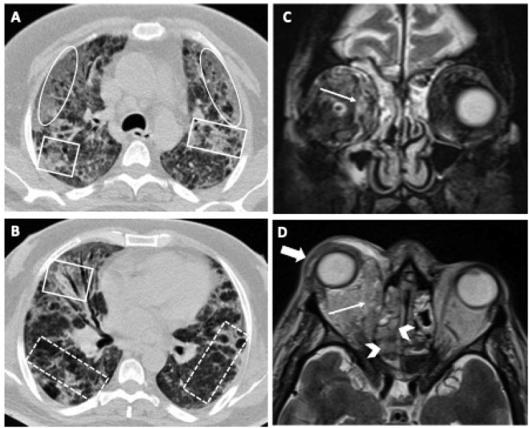
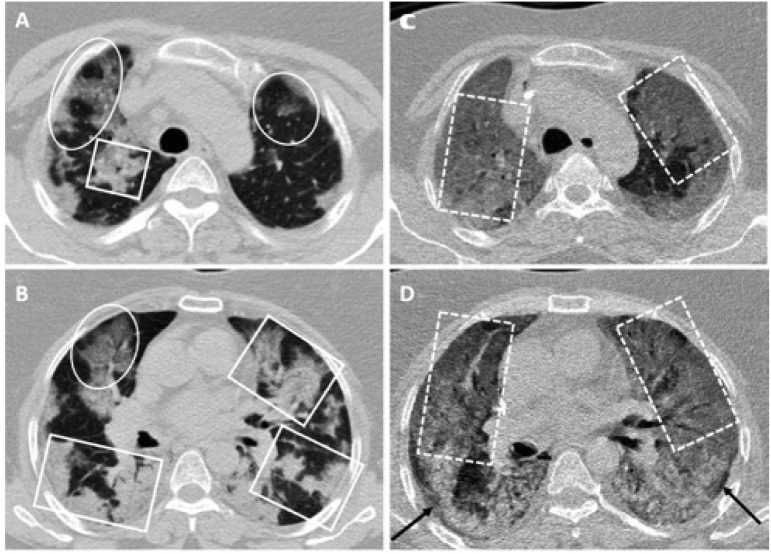
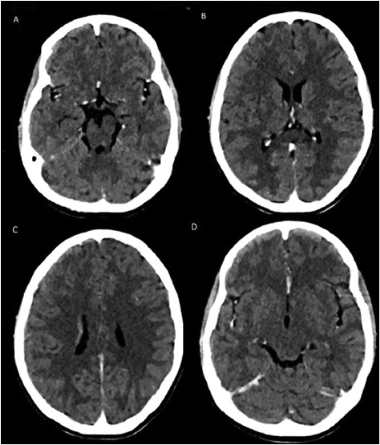
Globally, many hospitalized COVID-19 patients can experience an unexpected acute change in status, prompting rapid and expert clinical assessment. Superimposed infections can be a significant cause of clinical and radiologic deviations in this patient population, further worsening clinical outcome and muddling the differential diagnosis. As thrombotic, inflammatory, and medication-induced complications can also trigger an acute change in COVID-19 patient status, imaging early and often plays a vital role in distinguishing the cause of patient decline and monitoring patient outcome. While the common radiologic findings of COVID-19 infection are now widely reported, little is known about the clinical manifestations and imaging findings of superimposed infection. By discussing case studies of patients who developed bacterial, fungal, parasitic, and viral co-infections and identifying the most frequently reported imaging findings of superimposed infections, physicians will be more familiar with common infectious presentations and initiate a directed workup sooner. Ultimately, any abrupt changes in the expected COVID-19 imaging presentation, such as the presence of new consolidations or cavitation, should prompt further workup to exclude superimposed opportunistic infection.

Keywords: COVID-19; Co-infection; Opportunistic infection; Superimposed infection.


Conflict of interest statement

Declaration of competing interest The authors declare they have no conflict of interest in this study.

Figures

Fig. 1
Fig. 1
A 62-year-old male presented with acute hypoxemia with positive PCR for COVID -19. Initial frontal radiograph (A) shows multifocal peripheral predominant patchy opacities, which was confirmed on axial CT images (B and C) as basilar and peripheral predominant ground glass opacities with early superimposed consolidations (arrows), commonly seen in COVID-19 pneumonia. He was placed on supplemental oxygen with nasal cannula and started on hydroxychloroquine. On the 2nd day of admission, severe acute chest pain, hypotension and worsening respiratory failure required transfer to ICU, intubation and mechanical ventilation with initiation of empiric therapy for bacterial pneumonia. Further work up confirmed myocardial infarction and emergent left circumflex coronary artery stenting was performed. Over next week, his oxygen demands increased and follow up CT (D and E) demonstrated more confluent consolidations bilaterally along with new pleural effusions (circles). Subsequent, bronchial lavage/pleural cultures came consistent with Pseudomonas aeruginosa and antibiotic therapy was changed to Meropenem. Patient subsequently improved and discharged two weeks later to rehab on oxygen support.
Fig. 2
Fig. 2
SARS-CoV-2 and Legionella Co-infection. Frontal chest radiograph (A) of a patient with severe acute respiratory derangement and confirmed SARS-CoV-2 and Legionella co-infection, reveals no significant abnormality. However, on axial CT scan images (B, C and D) obtained on day 7 of illness, there are bilateral subpleural predominant patchy ground-glass opacities (arrows), which are typical for COVID-19 and no definite imaging clues to suggest co-infection.
Fig. 3
Fig. 3
Bacterial (Klebsiella) superinfection in COVID-19. A 54-year-old female patient with RT-PCR positive COVID-19 pneumonia (A, B). First CT scan upon admission (day 1) shows multifocal patchy ground-glass opacities (circles) and consolidations (boxes), mostly in left lung. He underwent treatment by antiviral agent and corticosteroids and improved significantly. On day 11, he underwent a second CT scan (C, D) due to persistent low O2 saturation without fever or cough which demonstrated new air space consolidations in right upper and right lower lobes (dotted boxes) with a decrease in ground-glass opacites. A bulging fissure sign is also noted (arrow in C). Culture of bronchoalveolar lavage confirmed superimposed Klebsiella infection.
Fig. 4
Fig. 4
SARS-CoV-2 and E.Coli Co-infection. A 62-year-old healthy male with RT-PCR positive COVID-19 pneumonia. Comparison of chest X-ray changes on day 1 (left) and day 5 (right) of presentation. CXR on day 5 (right) indicated a focal consolidation in the left upper lung, bilateral airspace opacities, and low lung volumes.
Fig. 5
Fig. 5
Aspergillosis superinfection in COVID-19. A 42-year-old female patient with newly diagnosed Acute Myeloid leukemia who recently underwent standard combination chemotherapy. Axial chest CT images (A, B) on day 8 revealed multiple vessel-related nodular opacities with ground glass halo with central and peripheral distribution(arrows in A and B) with small bilateral pleural effusion (stars in A).The imaging findings are more typical for an invasive fungal infection and atypical for COVID-19. She tested positive for SARS-CoV-2 and Aspergillus fumigatus.
Fig. 6
Fig. 6
Mixed fungal and bacterial co-infection in COVID-19 pneumonia. Initial axial chest CT images (A, B) on first day of admission reveal bilateral multifocal patchy ground-glass opacities (circles) and consolidations (arrows) with peripheral predominance. Following standard treatment by remdesivir and corticosteroid and partial improvement he developed fever, dyspnea and low O2 saturation. Second CT scan images ((C, D)) on day 14 show multifocal new parenchymal cavities in upper and lower lobes along with coarse reticular pattern. Air-fluid level is also seen in left lower lobe cavities. The imaging findings are suggestive of necrotizing pneumonia. He underwent bronchoscopy and culture of bronchoalveolar lavage revealed mixed fungal (aspergillosis) and gram-negative (Klebsiella) infection.
Fig. 7
Fig. 7
Superimposed Histoplasmosis infection on COVID-19. A 43-year-old woman with a 21-year history of HIV infection and poor adherence to antiretroviral treatment recently tested positive for COVID-19. Initial chest CT axial image (A) shows bilateral ground-glass opacities, diffuse bronchial wall thickening (arrow), and multiple centrilobular nodules (circles). 8 days later, Chest (C) and abdominal CT (D) were re-ordered due to worsening symptoms, which show worsening of pulmonary micro nodularity and ground glass opacification. Hepatosplenomegaly with numerous splenic hypodense nodules (circle in Fig. 7D) are visualized, concerning for disseminated granulomatous infection. Patient tested positive for urinary H. capsulatum antigen.
Fig. 8
Fig. 8
Rhino-orbital Mucormycosis in COVID-19. A 55-year-old male patient with recent RT-PCR positive COVID-19 pneumonia and history of longstanding poorly controlled diabetes mellitus. Chest CT scan (A, B) at the second week of infection upon ICU admission show extensive bilateral confluent consolidations (boxes), ground-glass opacities (ovals) and coarse reticular pattern (dotted boxes) associated with interstitial septal thickening suggestive for subacute phase of COVID-19 infection. He underwent standard of care treatment with remdesivir and corticosteroid. On day 4 of admission, he developed right sided proptosis, chemosis, and periorbital edema. Coronal (C) and axial (D) T2-weighted images from orbital MRI show opacification of bilateral frontal and ethmoidal sinuses as well as right maxillary sinus with areas of internal relative T2-weighted hypointense signal (arrowheads), typical for mucormycosis. An ill-defined lesion is depicted in medial right orbit (thin arrows) involving extra and intraconal spaces leading to proptosis of globe (thick arrow). Paranasal sinus culture confirmed rhino-orbital mucormycosis. He eventually died during admission following intracranial spread of fungal infection.
Fig. 9
Fig. 9
Pneumocystis pneumonia (PCP) in COVID-19. A 57-year-old female patient with RT-PCR positive COVID-19 pneumonia. Axial CT scan images (A, B) on day 1 show bilateral multifocal consolidations (boxes) and patchy ground-glass opacities (circles) with dominant peripheral distribution in the upper lobes and the superior segments of lower lobes. The patient did not respond well to routine standard of care treatment and corticosteroids were continued. Due to persistent hypoxemia and respiratory distress second CT was obtained on day 18 of admission in ICU (C, D), which shows new/worsened extensive bilateral confluent ground-glass opacities and fine reticular pattern (dotted boxes) with previously noted consolidations not clearly visualized. Relative subpleural sparing is also noted in the superior segment of lower lobes (arrows). Diagnostic bronchoscopy was canceled due to respiratory distress. Considering the new imaging findings, he underwent treatment trial for pneumocystis pneumonia (PCP) and dramatically improved and discharged.
Fig. 10
Fig. 10
COVID-19-associated Encephalitis with Dengue Shock Syndrome. A 14-year- old female presented with high-grade fever, headache and vomiting and respira- tory distress and hypotensive shock. Nasopharyngeal PCR testing for SARS-CoV-2 and serum dengue NS1 antigen were tested positive. Contrast-enhanced axial CT images showing ill-defined marked hypodensities in Mid-brain, thalamus, corpus callosum and bilateral periventricular area, basal ganglia and bilateral frontal lobes. Given the clinical context, lab testing results and patient's demographics (resident of an endemic for Dengue fever), the image findings are suggestive of viral encephalitis.

Similar articles

Cited by

References

    1. https://www.worldometers.info/coronavirus
    1. Cozzi D., Albanesi M., Cavigli E. Chest X-ray in new Coronavirus Disease 2019 (COVID-19) infection: findings and correlation with clinical outcome. Radiol. Med. 2020;125:730–737. - PMC - PubMed
    1. Kooraki S., Hosseiny M., Myers L., Gholamrezanezhad A. Coronavirus (COVID-19) outbreak: what the department of radiology should know. J Am Coll Radiol. 2020 Apr;17(4):447–451.. doi: 10.1016/j.jacr.2020.02.008. Epub 2020 Feb 19. PMID: 32092296; PMCID: PMC7102595. - DOI - PMC - PubMed
    1. Fields B.K.K., Demirjian N.L., Dadgar H., Gholamrezanezhad A. Imaging of COVID-19: CT, MRI, and PET. Semin Nucl Med. 2021 Jul;51(4):312–320. doi: 10.1053/j.semnuclmed.2020.11.003. Epub 2020 Nov 30. PMID: 33288215; PMCID: PMC7703471. - DOI - PMC - PubMed
    1. Hosseiny M., Kooraki S., Gholamrezanezhad A., Reddy S., Myers L. Radiology perspective of Coronavirus Disease 2019 (COVID-19): lessons from severe acute respiratory syndrome and middle east respiratory syndrome. AJR Am J Roentgenol. 2020 May;214(5):1078–1082. doi: 10.2214/AJR.20.22969. Epub 2020 Feb 28. PMID: 32108495. - DOI - PubMed