. 2022 Oct:171:117-132.
doi: 10.1016/j.yjmcc.2022.08.003. Epub 2022 Aug 22.

Loss of Acta2 in cardiac fibroblasts does not prevent the myofibroblast differentiation or affect the cardiac repair after myocardial infarction

Affiliations

Loss of Acta2 in cardiac fibroblasts does not prevent the myofibroblast differentiation or affect the cardiac repair after myocardial infarction

Yuxia Li et al. J Mol Cell Cardiol. 2022 Oct.

Abstract

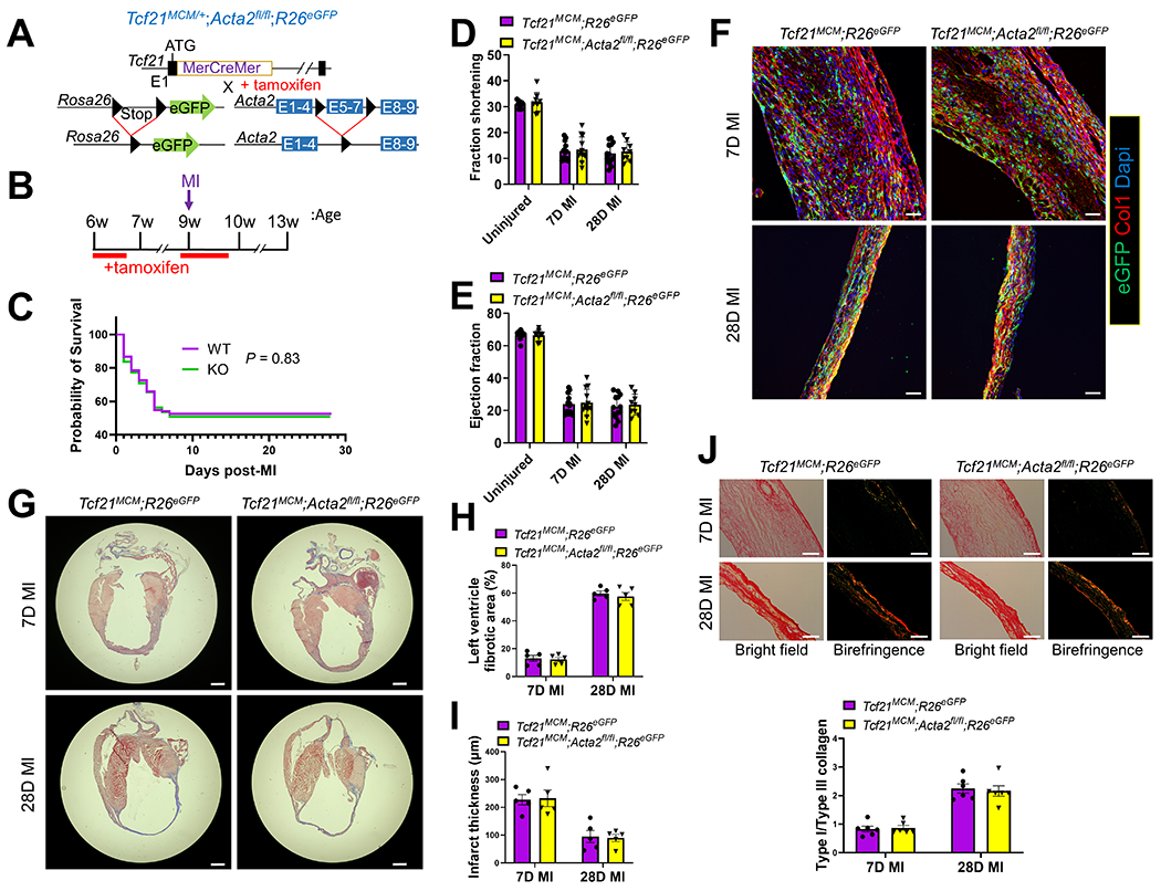
In response to myocardial infarction (MI), quiescent cardiac fibroblasts differentiate into myofibroblasts mediating tissue repair. One of the most widely accepted markers of myofibroblast differentiation is the expression of Acta2 which encodes smooth muscle alpha-actin (SMαA) that is assembled into stress fibers. However, the requirement of Acta2/SMαA in the myofibroblast differentiation of cardiac fibroblasts and its role in post-MI cardiac repair remained unknown. To answer these questions, we generated a tamoxifen-inducible cardiac fibroblast-specific Acta2 knockout mouse line. Surprisingly, mice that lacked Acta2 in cardiac fibroblasts had a normal post-MI survival rate. Moreover, Acta2 deletion did not affect the function or histology of infarcted hearts. No difference was detected in the proliferation, migration, or contractility between WT and Acta2-null cardiac myofibroblasts. Acta2-null cardiac myofibroblasts had a normal total filamentous actin level and total actin level. Acta2 deletion caused a significant compensatory increase in the transcription level of non-Acta2 actin isoforms, especially Actg2 and Acta1. Moreover, in myofibroblasts, the transcription levels of cytoplasmic actin isoforms were significantly higher than those of muscle actin isoforms. In addition, we found that myocardin-related transcription factor-A is critical for myofibroblast differentiation but is not required for the compensatory effects of non-Acta2 isoforms. In conclusion, the Acta2 deletion does not prevent the myofibroblast differentiation of cardiac fibroblasts or affect the post-MI cardiac repair, and the increased expression and stress fiber formation of non-SMαA actin isoforms and the functional redundancy between actin isoforms are able to compensate for the loss of Acta2 in cardiac myofibroblasts.

Keywords: Actin; Cardiac fibroblast; Myocardial infarction; Stress fiber.


Conflict of interest statement

Declaration of Competing Interest None.

Figures

Figure 1.
Figure 1.
Cardiac fibroblast-specific deletion of Acta2 does not affect the post-MI survival or cardiac function of mice. (A) Schematic of the generation of Tcf21MCM/+;Acta2fl/fl;R26eGFP mice. In these mice, a tamoxifen-regulated MerCreMer cDNA cassette inserted into exon 1 (E1) enables the tamoxifen-induced deletion of the loxP site-flanked stop cassette upstream of eGFP inserted into the R26 locus and the deletion of the loxP site-flanked exons 5-7 (E5-7) of Acta2 only in cells expressing Tcf21. (B) Experimental scheme of tamoxifen treatment and surgical procedure to induce MI. (C) Survival curves showing the survival rate of Tcf21MCM/+;R26eGFP and Tcf21MCM/+;Acta2fl/fl;R26eGFP mice after MI. n=135 (Tcf21MCM/+;R26eGFP); n=110 (Tcf21MCM/+;Acta2fl/fl;R26eGFP). (D-E) Fraction shortening (D) and ejection fraction (E) of Tcf21MCM/+;R26eGFP and Tcf21MCM/+;Acta2fl/fl;R26eGFP mice without MI and at 7 and 28 days after MI. n=9 for uninjured mice; n=13 for Tcf21MCM/+;R26eGFP; mice at 7 and 28 days after MI; n=12 for for Tcf21MCM/+;Acta2fl/fl;R26eGFP mice 7 days after MI; n=9 for Tcf21MCM/+;Acta2fl/fl;R26eGFP mice 28 days after MI. (F-I) Heart samples were collected from tamoxifen-treated Tcf21MCM/+;R26eGFP and Tcf21MCM/+;Acta2fl/fl;R26eGFP mice at 7 and 28 days after MI. (F) Representative IHC images from 5 analyzed hearts per group show the presence of eGFP+ Tcf21 lineage-traced cardiac fibroblasts and the expression of type I collagen (Col1). Nuclei are shown with Dapi. Scale bar: 50 μm. (G) Representative trichrome staining images from 5 analyzed hearts per group showing the dilation of the left ventricle after MI and collagen deposition in the infarct area. Scale bar: 1 mm. (H-I) Percentage of the fibrotic area in the left ventricle (H) and average left ventricle thickness (I) determined by analysis of trichrome staining images using ImageJ. n=5 for each group. (J) Representative picrosirius red staining images from 6 analyzed hearts per group showing the collagen maturation (type I/III collagen ratio) in the infarct area. n=6. Scale bar: 50 μm.
Figure 2.
Figure 2.
Cardiac fibroblast-specific deletion of Acta2 does not alter cardiac fibroblast proliferation after MI. (A-B) Tamoxifen-treated Tcf21MCM/+;R26eGFP and Tcf21MCM/+;Acta2fl/fl;R26eGFP mice were subjected to MI and EdU based proliferation assays at 3 days and 7 days post-MI. IHC was performed to identify and quantify Tcf21 lineage-traced (eGFP+) cardiac fibroblasts that are positive for SMαA, Ki67, or EdU in the infarct region at 3 days (A) and 7 days (B) after MI. n=3 for each group. Scale bar: 20 μm. (C) ICC was performed to identify the expression of SMαA and Ki67 in WT and Acta2-null cardiac myofibroblasts. Nuclei are shown with Dapi. n=4 for each group. Scale bar: 100 μm. (D) Cardiac fibroblasts isolated from R26tdTomato (WT) and Acta2fl/fl;R26eGFP mice were transduced with Adeno-Cre and co-cultured at a 1:1 ratio. The ratio between WT and Acta2-null cardiac fibroblasts was calculated again after 48 hours of co-culture in the presence of TGFβ. n=3 for each group. Scale bar: 250 nm. **P < 0.01; ***P < 0.0001.
Figure 3.
Figure 3.
Deletion of Acta2 does not affect the motility, contractility, and matrix stabilization ability of cardiac fibroblasts. (A) WT (tdTomato+) and Acta2-null (GFP+) cardiac myofibroblasts were seeded onto the same wells of 96-well plates at a 1:1 ratio. The migration of cells into the center of wells was quantified every day. n=3 for each group. Scale bar: 300 μm. (B-C) WT and Acta2-null cardiac myofibroblasts were mixed with collagen gel and poured onto 24 well plates. (B) Gels were released from wells after 12 hours of incubation. Images and quantification show the degree of contraction of gels 48 hours after the release of gels. n=3 for each group. (C) The storage moduli of cell-free gels and cell-laden gels 24 hours after incubation were measured using a rheometer. n=5 for each group.
Figure 4.
Figure 4.
Deletion of Acta2 does not prevent the myofibroblast differentiation of cardiac fibroblasts. (A) WT and Acta2-null cardiac myofibroblasts were subjected to ICC to identify the expression of SMαA and presence of F-actin using an anti-SMαA antibody and phalloidin, respectively. Nuclei are shown with Dapi. Scale bar: 100 μm. The fluorescence intensity was determined using ImageJ. n=3 for each group. **P < 0.01. (B) Cardiac fibroblasts isolated from tamoxifen-treated Tcf21MCM/+;R26eGFP and Tcf21MCM/+;Acta2fl/fl;R26eGFP mice were treated with TGFβ for 2 days and subjected to ICC to identify the expression of SMαA and presence of F-actin. Nuclei are shown with Dapi. Images represent 3 independent replications. Scale bar: 40 μm. (C) IHC was performed to identify Tcf21 lineage-traced (eGFP+) cardiac fibroblasts that are positive for SMαA or/and phalloidin in Tcf21MCM/+;R26eGFP and Tcf21MCM/+;Acta2fl/fl;R26eGFP mice at post-MI day 7. Nuclei are shown with Dapi. Images represent 3 analyzed hearts per group. Scale bar: 20 μm. (D) WT and Acta2-null cardiac myofibroblasts were subjected to ICC to identify the expression of SMαA and vinculin using specific antibodies. Nuclei are shown with Dapi. n=4 for each group. Scale bar: 40 μm. The average area of focal adhesion was measured using ImageJ. (E-F) Western blot (E) and ICC (F) were performed to quantify SMαA and total actin protein levels in WT and Acta2-null cardiac myofibroblasts using an anti-SMαA antibody and a pan-actin antibody, respectively. Scale bar: 100 μm. n=3 (western blot) or n=4 (ICC) for each group; **P < 0.01.
Figure 5.
Figure 5.
The compensatory effects of non-SMαA actin isoforms in Acta2-null cardiac myofibroblasts. (A) Tcf21 lineage-traced cardiac fibroblasts sorted from uninjured hearts and the infarct region at 3 days and 7 days after MI were subjected to RNAseq. The normalized transcription levels (TPM) of Acta2, Acta1, Actb, Actc1, Actg1, and Actg2 are shown. n=2 for each group. *P < 0.05; ***P < 0.0001 vs uninjured. (B) The transcription levels of Acta2, Acta1, Actb, Actc1, Actg1, and Actg2 in cultured WT and Acta2-null cardiac myofibroblasts were revealed by RNAseq. n=2 for each group. **P < 0.01. (C) Tcf21 lineage-traced cardiac fibroblasts were sorted from the infarct scar of Tcf21MCM/+;R26eGFP and Tcf21MCM/+;Acta2fl/fl;R26eGFP mice at post-MI day 7. The expression level of Acta2, Acta1, Actg1, Actg2, Col1a1, Col3a1, and Postn were revealed by realtime PCR. n=4 for each group. *P < 0.05; **P < 0.01. (D-E) WT and Acta2-null cardiac myofibroblasts were subjected to ICC to identify the protein level of SMαA, the combined protein level of SkMαA and CMαA, and the combined protein level of SMαA and SMγA using specific antibodies (D). Nuclei are shown with Dapi. Higher-magnification images highlight the areas marked by solid-line boxes (color-coded). Scale bar: 100 μm. The fluorescence intensity was measured using Image J (E). n=3 for WT; n=4 for Acta2-null. **P < 0.01; ***P < 0.0001. (F-G) Western blot (F) and ICC (G) were performed to quantify SMαA, CyβA, and CyγA protein levels in WT and Acta2-null cardiac myofibroblasts using specific antibodies. Higher-magnification images highlight the areas marked by yellow solid-line boxes. n=3 for each group (F). Images represent 3 independent replications (G). Scale bar: 100 μm.
Figure 6.
Figure 6.
Knocking down of Mrtfa reduces the expression of muscle actin isoforms but does not affect the compensatory effects of non-SMαA actin isoforms in Acta2-null cardiac myofibroblasts. WT and Acta2-null cardiac fibroblasts were treated with Lenti-GFP or Lenti-shMrtfa and induced for myofibroblast differentiation using TGFβ for 2 days. (A-C) ICC was performed to identify the expression of SMαA (A-B), total F-actin level (by phalloidin) (A), and total actin level (B). Scale bar: 40 μm (A), 100 μm (B). The fluorescence intensity was measured using Image J (C). n=4 for each group. Different letters indicate samples with significant differences (P < 0.05). (D) Realtime PCR was performed to identify the transcription levels of Acta2, Acta1, Actb, Actc1, Actg1, and Actg2. n=3 for each group. Different letters indicate samples with significant differences (P< 0.05). (E-G) ICC was performed to identify the expression of SMαA (F-G), SkMαA/CMαA (F), SMα/γA (F), CyβA (G), and CyγA (G). The fluorescence intensity was measured using Image J (E). Different letters indicate samples with significant differences (P < 0.05). n=4 for each group. Scale bar: 100 μm (F) and 40 μm (G).

Similar articles

Cited by

References

    1. Sheifer SE, Gersh BJ, Yanez ND 3rd, Ades PA, Burke GL, Manolio TA, Prevalence, predisposing factors, and prognosis of clinically unrecognized myocardial infarction in the elderly, J. Am. Coll. Cardiol 35(1) (2000) 119–26. - PubMed
    1. Yeh RW, Sidney S, Chandra M, Sorel M, Selby JV, Go AS, Population trends in the incidence and outcomes of acute myocardial infarction, N. Engl. J. Med 362(23) (2010) 2155–65. - PubMed
    1. Yellon DM, Hausenloy DJ, Myocardial reperfusion injury, New Engl. J. Med 357(11) (2007) 1121–1135. - PubMed
    1. Fu X, Khalil H, Kanisicak O, Boyer JG, Vagnozzi RJ, Maliken BD, Sargent MA, Prasad V, Valiente-Alandi I, Blaxall BC, Molkentin JD, Specialized fibroblast differentiated states underlie scar formation in the infarcted mouse heart, J. Clin. Invest 128(5) (2018) 2127–2143. - PMC - PubMed
    1. Davis J, Molkentin JD, Myofibroblasts: trust your heart and let fate decide, J. Mol. Cell. Cardiol 70 (2014) 9–18. - PMC - PubMed

Publication types