. 2022 Oct 30;434(20):167799.
doi: 10.1016/j.jmb.2022.167799. Epub 2022 Aug 22.

Terminase Subunits from the Pseudomonas-Phage E217

Affiliations

Terminase Subunits from the Pseudomonas-Phage E217

Ravi K Lokareddy et al. J Mol Biol. .

Abstract

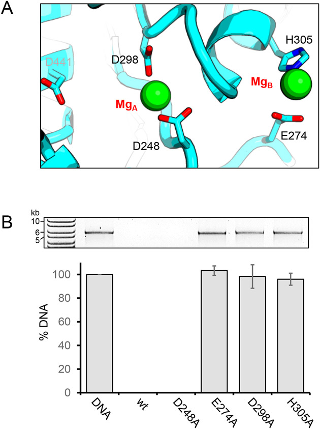
Pseudomonas phages are increasingly important biomedicines for phage therapy, but little is known about how these viruses package DNA. This paper explores the terminase subunits from the Myoviridae E217, a Pseudomonas-phage used in an experimental cocktail to eradicate P. aeruginosa in vitro and in animal models. We identified the large (TerL) and small (TerS) terminase subunits in two genes ∼58 kbs away from each other in the E217 genome. TerL presents a classical two-domain architecture, consisting of an N-terminal ATPase and C-terminal nuclease domain arranged into a bean-shaped tertiary structure. A 2.05 Å crystal structure of the C-terminal domain revealed an RNase H-like fold with two magnesium ions in the nuclease active site. Mutations in TerL residues involved in magnesium coordination had a dominant-negative effect on phage growth. However, the two ions identified in the active site were too far from each other to promote two-metal-ion catalysis, suggesting a conformational change is required for nuclease activity. We also determined a 3.38 Å cryo-EM reconstruction of E217 TerS that revealed a ring-like decamer, departing from the most common nonameric quaternary structure observed thus far. E217 TerS contains both N-terminal helix-turn-helix motifs enriched in basic residues and a central channel lined with basic residues large enough to accommodate double-stranded DNA. Overexpression of TerS caused a more than a 4-fold reduction of E217 burst size, suggesting a catalytic amount of the protein is required for packaging. Together, these data expand the molecular repertoire of viral terminase subunits to Pseudomonas-phages used for phage therapy.

Keywords: Pseudomonas-phages; bacteriophage E217; large terminase; small terminase; viral genome-packaging motor.


Conflict of interest statement

Declaration of interests The authors declare that they have no known competing financial interests or personal relationships that could have appeared to influence the work reported in this paper.

Figures

Figure 1.
Figure 1.. Crystal structure of E217 TerL nuclease domain at 2.05 Å.
(A) Schematic diagram of E217 TerL showing N-terminal ATPase and C-terminal nuclease domains. (B) A time course of proteolytic cleavage of purified E217 TerL in the presence of chymotrypsin yields a stable nuclease core. (C) Crystal structure of nuclease domain residues 206-453 refined at 2.05 Å resolution. Two Mg2+ ions (MgA and MgB) identified in the electron density are shown as green spheres. (D) Magnified view of the β-hairpin residues 330-357 that adopt different conformations in the two chains in the asymmetric unit (colored in cyan and gray). The maximum displacement between chains A and B is ~5Å.
Figure 2.
Figure 2.. Solution structure of E217 full-length TerL.
(A) Left, experimental scattering profile of the TerL (black trace) overlaid with Rg distribution across the scattering peak (red circles). Center, Guinier region of the intensity I(q) to the scattering vector (q2). The qmax(Rg) cut-off was 1.3. Right, P(r) function with Dmax of 84.5 Å. (B) Model of TerL fit within the electron density generated by DENSS. TerL N-terminal ATPase and C-terminal nuclease domain are shown in yellow and cyan, respectively. (C) The comparison of the scattering profile predicted for the model to the empirical scattering of the complex produced a χ2 value of 1.03.
Figure 3.
Figure 3.. Critical residues in TerL nuclease active site.
(A) Magnified view of the magnesium ions found in the nuclease active site. MgA is bound by D248 and D298, while E274 and H305 chelate MgB. Another residue, D441, appears too far to contact MgA in the conformation seen in crystals. (B) In vitro nuclease assay. Linearized pET28-vector was incubated with TerL and relative mutants for 1h at 37 °C. The reaction mixture was separated on 0.8% agarose and DNA visualized with ethidium bromide. Bars represent average with standard deviation (N= 3).
Figure 4.
Figure 4.. In vivo burst assay.
PAO1 cultures overexpressing the indicated TerL or TerS variants were infected with E217. Bars represent the average with standard deviation (N≥3) of the number of infectious phage particles released by each infected cell. Wt and D248A TerL did not interfere with E217 growth, whereas E274A, D298A, and H305A TerL-reduced the quantity of phage progeny relative to that produced by PAO1 with the empty vector (vector). Similarly, for TerS mutants, both wt TerS and the double mutant (K27A/R28A) dramatically interfered with E217 progeny. Significance was evaluated with t-test with respect to E217 burst size in PAO1 carrying the empty vector. ns, not significant; **, P≤0.01.
Figure 5.
Figure 5.. Cryo-EM single-particle analysis of E217 TerS.
(A) Representative 2D-class averages of the purified E217 TerS. The red asterisk indicates the fuzzy density emanating from the N-termini. (B) Representative sharpened electron density of E217 TerS overlaid with helix H5 residues A82 – P105. The density was contoured at 1σ and 3.4 Å resolution at FSC = 0.143. (C) Ribbon diagram for E217 TerS quaternary structure in top and side views. A semi-transparent solvent surface is overlaid on the ribbon diagram. The zoom-in panels show a magnified view of the central channel that is 22 Å in diameter between K108 of juxtaposed subunits.
Figure 6.
Figure 6.. E217 TerS tertiary structure.
(A) Schematic diagram of E217 TerS showing folded α-helices visible in the cryo-EM reconstruction and invisible N- and C-terminal moieties as rectangular boxes and gray lines, respectively. Alfafold predicted α-helix spanning C-terminal residues 140-163 is shown as an insert (with acidic residues colored in red). (B) Ribbon diagram of the TerS protomer modeled in the cryo-EM reconstruction with α-helices shown as cylinders. Putative residues in H1-H3 involved in DNA-binding are shown as sticks.
Figure 7.
Figure 7.. E217 TerS residues putatively involved in DNA-binding.
Coulombic electrostatic potential surface of E217 TerS calculated using ChimeraX [78]. (A) A bottom view of TerS reveals basic residues mainly projecting outward and lining the central channel. (B) A section through the central channel reveals two basic residues: K108 and K117. (C) The channel diameter and shape were calculated using MOLE 2.5 (mole.upol.cz).

Similar articles

Cited by

References

    1. Sun S, Rao VB, Rossmann MG. Genome packaging in viruses. Curr Opin Struct Biol. 2010;20:114–20. - PMC - PubMed
    1. Rao VB, Feiss M. The bacteriophage DNA packaging motor. Annu Rev Genet. 2008;42:647–81. - PubMed
    1. Casjens SR. The DNA-packaging nanomotor of tailed bacteriophages. Nat Rev Microbiol. 2011;9:647–57. - PubMed
    1. Guo P, Zhao Z, Haak J, Wang S, Wu D, Meng B, et al. Common mechanisms of DNA translocation motors in bacteria and viruses using one-way revolution mechanism without rotation. Biotechnol Adv. 2014;32:853–72. - PMC - PubMed
    1. Fuller DN, Raymer DM, Kottadiel VI, Rao VB, Smith DE. Single phage T4 DNA packaging motors exhibit large force generation, high velocity, and dynamic variability. Proc Natl Acad Sci U S A. 2007;104:16868–73. - PMC - PubMed

Publication types

MeSH terms