Review
. 2023 Jan 15;435(1):167800.
doi: 10.1016/j.jmb.2022.167800. Epub 2022 Aug 22.

Physical-Chemical Regulation of Membrane Receptors Dynamics in Viral Invasion and Immune Defense

Affiliations
Review

Physical-Chemical Regulation of Membrane Receptors Dynamics in Viral Invasion and Immune Defense

Rui Qin et al. J Mol Biol. .

Abstract

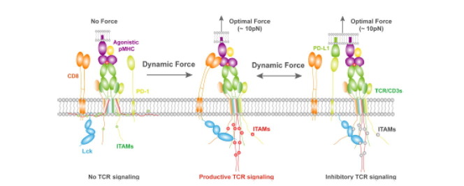
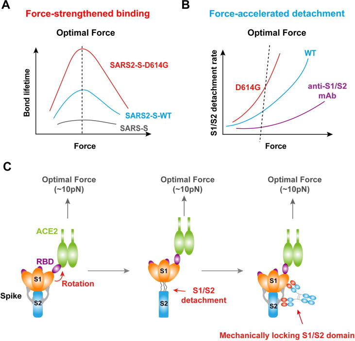
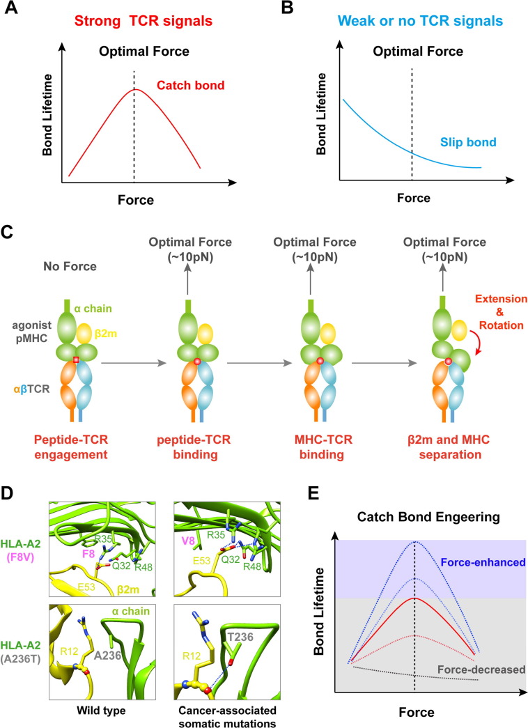
Mechanical cues dynamically regulate membrane receptors functions to trigger various physiological and pathological processes from viral invasion to immune defense. These cues mainly include various types of dynamic mechanical forces and the spatial confinement of plasma membrane. However, the molecular mechanisms of how they couple with biochemical cues in regulating membrane receptors functions still remain mysterious. Here, we review recent advances in methodologies of single-molecule biomechanical techniques and in novel biomechanical regulatory mechanisms of critical ligand recognition of viral and immune receptors including SARS-CoV-2 spike protein, T cell receptor (TCR) and other co-stimulatory immune receptors. Furthermore, we provide our perspectives of the general principle of how force-dependent kinetics determine the dynamic functions of membrane receptors and of biomechanical-mechanism-driven SARS-CoV-2 neutralizing antibody design and TCR engineering for T-cell-based therapies.

Keywords: immune defense; mechanical force; membrane receptors dynamics; spatial confinement of plasma membrane; viral invasion.


Conflict of interest statement

Declaration of Interest Authors declare that they have no competing interests.

Figures

None
Graphical abstract
Figure 1
Figure 1
Mechanical forces strengthen the spike/ACE2 binding and accelerate S1/S2 detachment, providing novel intervention strategy. (A) Schematics of force-dependent bond lifetimes of the interactions of host ACE2 receptors interacting with SARS2-S-D614G (red), SARS2-S-WT (blue) and SARS-S (gray), respectively. (B) S1/S2 detachment rate versus force curves of SARS2-S-D614G (red), SARS2-S-WT (blue) and SARS2-S-WT in the presence of neutralizing antibody targeting S1/S2 (purple), respectively. (C) The dynamic structural model of force-regulated SARS2-S/ACE2 binding and conformational change of SARS2-S, force-induced S1/S2 detachment, and S1/S2-locking antibodies to impede SARS2-S/ACE2 binding.
Figure 2
Figure 2
Force-induced conformational changes regulate catch bond formation between TCRs and pMHCs. (A, B) Bond lifetime versus force curves showing that TCR forms catch-slip bonds with agonists pMHC (red) (A) but slip bonds with antagonists pMHC (blue) (B). (C) Dynamic sequential steps in the mechanical regulation of pMHC conformation between TCR and agonistic pMHC. (D) Cancer-associated somatic mutations (A236T and F8V in HLA-A2) induce new H-bonds between α and β2 m subunits. H-bonds are indicated as blue dashed lines. (E) Mechanical forces modulate enhancement or reduction of the catch bond between TCRs and pMHCs.
Figure 3
Figure 3
A dynamic model of mechano-chemical coupling for receptor-ligand interactions and transmembrane signaling. When TCR recognizes agonistic pMHC, force induces conformational changes in pMHC and the extracellular domains of TCR, which sequentially propagates across the cell membrane to induce intracellular kinase activation (Lck) and structural changes in the cytoplasmic domains (CD3). Meanwhile, CD8-associated Lck binds to phosphorylated CD3 ITAMs inside the cell membrane and to TCR-CD3 complex outside the cell membrane, forming a positive feedback mechanical loop that amplifies productive TCR signaling. However, the TCR-CD8 cooperative binding of pMHC is disrupted in the presence of PD-1, manifesting a negative or inhibitory cooperativity.

Similar articles

Cited by

References

    1. Yin H., Flynn A.D. Drugging Membrane Protein Interactions. Annu Rev Biomed Eng. 2015;18:1–26. doi: 10.1146/annurev-bioeng-092115-025322. - DOI - PMC - PubMed
    1. Hu W., Zhang Y., Fei P., Zhang T., Yao D., Gao Y., Liu J., Chen H., et al. Mechanical activation of spike fosters SARS-CoV-2 viral infection. Cell Res. 2021:1–14. doi: 10.1038/s41422-021-00558-x. - DOI - PMC - PubMed
    1. Zamoyska R. CD4 and CD8: modulators of T-cell receptor recognition of antigen and of immune responses? Curr Opin Immunol. 1998;10:82–87. doi: 10.1016/s0952-7915(98)80036-8. - DOI - PubMed
    1. Mørch A.M., Bálint Š., Santos A.M., Davis S.J., Dustin M.L. Coreceptors and TCR Signaling – the Strong and the Weak of It. Front Cell Dev Biol. 2020:1–13. https://www.frontiersin.org/ - PMC - PubMed
    1. He J., Xiong X., Yang H., Li D., Liu X., Li S., Liao S., Chen S., et al. Defined tumor antigen-specific T cells potentiate personalized TCR-T cell therapy and prediction of immunotherapy response. Cell Res. 2022:1–13. doi: 10.1038/s41422-022-00627-9. - DOI - PMC - PubMed

Publication types

Substances

LinkOut - more resources