. 2022 Aug 25;13(1):5004.
doi: 10.1038/s41467-022-32688-0.

Designed peptides as nanomolar cross-amyloid inhibitors acting via supramolecular nanofiber co-assembly

Affiliations

Designed peptides as nanomolar cross-amyloid inhibitors acting via supramolecular nanofiber co-assembly

Karin Taş et al. Nat Commun. .

Abstract

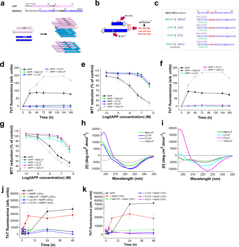
Amyloid self-assembly is linked to numerous devastating cell-degenerative diseases. However, designing inhibitors of this pathogenic process remains a major challenge. Cross-interactions between amyloid-β peptide (Aβ) and islet amyloid polypeptide (IAPP), key polypeptides of Alzheimer's disease (AD) and type 2 diabetes (T2D), have been suggested to link AD with T2D pathogenesis. Here, we show that constrained peptides designed to mimic the Aβ amyloid core (ACMs) are nanomolar cross-amyloid inhibitors of both IAPP and Aβ42 and effectively suppress reciprocal cross-seeding. Remarkably, ACMs act by co-assembling with IAPP or Aβ42 into amyloid fibril-resembling but non-toxic nanofibers and their highly ordered superstructures. Co-assembled nanofibers exhibit various potentially beneficial features including thermolability, proteolytic degradability, and effective cellular clearance which are reminiscent of labile/reversible functional amyloids. ACMs are thus promising leads for potent anti-amyloid drugs in both T2D and AD while the supramolecular nanofiber co-assemblies should inform the design of novel functional (hetero-)amyloid-based nanomaterials for biomedical/biotechnological applications.


Conflict of interest statement

A.K., J.B., and K.T. are co-inventors of the European patent application 22 158 021.0 (applicant Technical University of Munich) (status: pending) related to ACMs, their hetero-assemblies, and potential biomedical applications. The remaining authors declare no competing interests.

Figures

Fig. 1
Fig. 1. ACM design concept, their effects on IAPP amyloid self-assembly and cytotoxicity, and ACM secondary structures.
a Sequences of IAPP and Aβ40(42), proposed models of fIAPP and fAβ40 folds, and hypothetical IAPP/Aβ40 “hetero-amyloids” (β-strands, pink or blue and underlined; “hot segments” of self-/cross-interactions, bold; loop residues, italics),,. b ACM inhibitor design strategy. Template Aβ(15–40) in a β-strand-loop-β-strand fold proposed for fAβ40 is modified via (a) N-methylations in Aβ(17–20), b substitution of Aβ(24-26) by hydrophobic tripeptides, and c Met35 substitution by Nle. c Sequences of the six ACMs and negative controls VGS-VF and VGS-LF (Supplementary Table 1). Each sequence corresponds to two different ACMs which contain the same LTS but a different couple of N-methylated residues (dashed boxes). Color code as in a; LTS and tripeptide VGS in red; green or violet for peptide names and corresponding N-methylated residues. d Nle3-VF, L3-VF, and F3-VF block IAPP amyloid self-assembly. Fibrillogenesis of IAPP (16.5 µM) alone or with ACMs or VGS-VF was assessed via ThT binding (IAPP/peptide 1/2) (means ± SD, n = 3 independent assays). e Nle3-VF, L3-VF, and F3-VF suppress the formation of toxic IAPP assemblies. Solutions of d (7-day-aged (VFS-VF 24 h)) added to RIN5fm cells; cell viability determined via MTT reduction (means ± SD, three independent assays, n = 3 technical replicates each). f Nle3-LF, L3-LF, and F3-LF block IAPP amyloid self-assembly. Assay as in d (IAPP/peptide 1/2 except L3-LF (1/2.5)) (means ± SD, three independent assays). g Nle3-LF, L3-LF, and F3-LF suppress the formation of toxic IAPP assemblies. Solutions of f (7-day-aged (VGS-LF 24 h)) added to RIN5fm cells; cell viability determined via MTT reduction (means ± SD, three independent assays, n = 3 technical replicates each). h, i Secondary structure of ACMs. Far-UV CD spectra of ACMs of d and f versus non-inhibitors (5 µM, pH 7.4). j ACMs inhibit seeding of IAPP by preformed fIAPP. Fibrillogenesis of IAPP (12 µM) without or with fIAPP seeds (10%) and seeded IAPP/ACM mixtures assessed via ThT binding (IAPP/ACM 1/2) (means ± SD, n = 9 (for IAPP alone) or 3 (for all other samples) independent assays). k ACMs inhibit fAβ42-mediated cross-seeding of IAPP. Fibrillogenesis of IAPP with and without fAβ42 seeds (10%) versus IAPP/ACM mixtures (IAPP 12 µM, IAPP/ACM 1/2) (means ± SD, n = 6 (for IAPP with or without seeds) or n = 3 (for all other samples) independent assays).
Fig. 2
Fig. 2. Nanomolar affinity IAPP/ACM interactions yield amyloid fibril-resembling but ThT-invisible nanofibers.
a Nanomolar affinity IAPP/ACM interactions as determined by fluorescence spectroscopy. Fluorescence spectra of Fluos-IAPP (5 nM) and its mixtures with Nle3-VF (pH 7.4) at indicated molar ratios (data from one representative binding assay from n = 3 independent assays). Inset, binding curve; data are means ± SD from n = 3 independent assays. b IAPP/ACM interactions result in hetero-dimers and medium-to-high MW hetero-assemblies. Characterization of IAPP/Nle3-VF hetero-complexes via cross-linking at different time points and NuPAGE and Western blot using anti-IAPP (left) or anti-Aβ (right) antibodies. IAPP/Nle3-VF mixtures (1/2; IAPP, 30 µM) were cross-linked with glutaraldehyde; the orange box indicates medium-to-high MW hetero-assemblies (major species); arrows indicate hetero-di-/-tri-/tetramers. Representative results from >3 independent assays. c Characterization of IAPP/Nle3-VF hetero-assemblies via size exclusion chromatography (SEC). Chromatograms of IAPP alone (16.5 µM) or its mixtures with Nle3-VF (1/2) at 0 h and at 96 h (mAU, milli-absorbance units). The black arrow indicates IAPP monomers and Nle3-VF dimers; blue arrows indicate IAPP/Nle3-VF hetero-dimers and medium-to-high MW hetero-assemblies. Similar results were found in two independent assays. d IAPP/Nle3-VF co-assemblies are more disordered than β-sheet-rich IAPP assemblies. Far-UV CD spectra of 7-day-aged IAPP (16.5 µM; pH 7.4) and its mixtures (1/2) with the Nle3-VF or VGS-VF (non-inhibitor) are shown; for comparison, the spectrum of freshly dissolved IAPP (0 h) is also shown. CD spectra are the average of three spectra of the same solution. e IAPP/Nle3-VF co-assembly blocks surface-exposure of hydrophobic clusters occurring at early steps of IAPP amyloid self-assembly as determined by anilinonaphthalene 8-sulfonate (ANS) binding. Fluorescence emission spectra of ANS alone/with IAPP (2 µM) (left) and of ANS alone/with IAPP/ACM (1/2) mixtures (right) (pH 7.4) were measured at various time points of self- or co-assembly as indicated (representative results from two independent assays). f IAPP/ACM interactions result in ThT-invisible fibrils of indistinguishable appearance to fIAPP by TEM. TEM images of 7-day-aged IAPP (16.5 µM) and its mixtures (1/2) with ACMs or VGS-VF (non-inhibitor) (solutions from Fig. 1d, f). Scale bars: 100 nm. Representative images from seven independent assays for fIAPP and the IAPP/Nle3-VF mixture and two to three similar independent assays for the other IAPP/ACM mixtures. g fIAPP and fibrils in IAPP/Nle3-VF mixture exhibit the amyloid cross-β structure signature. X-ray fiber diffraction patterns of fIAPP and fibrils present in aged IAPP/Nle3-VF mixture (1/2) showed major meridional and equatorial reflections at ~4.7 and ~10 Å. Data were representative of two independent experiments.
Fig. 3
Fig. 3. Evidence for supramolecular IAPP/ACM nanofiber co-assembly.
a Immunogold-TEM images of aged IAPP/Nle3-VF (1/2; IAPP, 16.5 µM) reveals fibrils which bind to both anti-fIAPP and anti-Aβ antibodies (IAPP, 5 nm gold; Nle3-VF, 10 nm gold). Scale bars: 100 nm. Representative images from four biologically independent samples. b Heteromeric nature of ThT-invisible fibrils in aged Biotin-IAPP/Nle3-VF (1/2; Biotin-IAPP, 16.5 µM) as assessed by a biotin pull-down assay. Components were revealed by WB with anti-Aβ (upper part) and anti-IAPP (lower part) antibodies; lane “Nle3-VF (control)”, Nle3-VF directly loaded onto the gel (without beads) (data representative from two independent assays). c STED images of supramolecular heteromeric nanofiber bundles in aged IAPP/Nle3-VF (1/2; IAPP(total), 16.5 µM) containing TAMRA-IAPP and Atto647-Nle3-VF (10%). Scale bars: 5 µm. Nanofiber assemblies are representative of 11 assemblies found in one sample; consistent with the results of 2PM examination of three biologically independent samples (see d). d 2PM images of nanofiber bundles in aged IAPP-containing TAMRA-IAPP (10%) (left), aged IAPP/Nle3-VF containing TAMRA-IAPP and Atto647N-Nle3-VF (10%) (middle), and aged IAPP/Nle3-VF containing TAMRA-IAPP and Fluos-Nle3-VF (10%) (right) (1/2; IAPP(total), 16.5 µM). Scale bars: 10 µm. Assemblies are representative of assemblies observed in three (IAPP/ACM) and four (IAPP) biologically independent samples examined by 2PM (IAPP/Nle3-VF) and by 2PM or CLSM & STED (IAPP/Nle3-VF), respectively. eh 2PM images of heteromeric fibrous superstructures in aged TAMRA-IAPP/Fluos-Nle3-VF (1/2) (TAMRA-IAPP 16.5 μM). Short colored arrows indicate nanofiber bundles parallel or intertwined (red, TAMRA-IAPP; green, Fluos-Nle3-VF) or overlaying (yellow); long white arrows indicate twists or wrapping. Scale bars: panel (e) 5 µm, f upper part, 5 μm and lower parts, 1 μm, g 50 μm, h 50 μm (insets 5 μm). Images are representative of three biologically independent samples. i 2PM image of a huge nanotube-like co-assembly found in aged TAMRA-IAPP/Fluos-Nle3-VF (1/2; TAMRA-IAPP, 16.5 μM) (upper panel) and 3D reconstruction of z-stacks (lower panel). Scale bars: 100 µm (inset 10 µm). Data were representative from three biologically independent samples. j FLIM-FRET analysis of TAMRA-IAPP/Fluos-Nle3-VF co-assembly of g indicates a very close (<5.5 nm) donor-acceptor proximity. Left panel, fluorescence decay curves (top) and lifetimes (bottom) of donor (Fluos-Nle3-VF) without or with acceptor (TAMRA-IAPP); a strong shift of donor lifetime in the presence of acceptor is observed. Middle panel/left side, FLIM image showing donor lifetime; lifetime range 0 ns (dark blue) to 3.5 ns (red); scale bar, 50 µm. Middle panel/right side, FLIM-FRET efficiency (%); efficiency range 0% (dark blue) to 80% (red); scale bar, 50 µm. Right panel, distributions donor lifetime (<1 ns) and FLIM-FRET efficiency (~55%). Data were representative of two independent experiments.
Fig. 4
Fig. 4. Mechanism of formation and properties of IAPP/ACM nanofiber co-assemblies.
a Evolution of hf-IAPP/ACM from amorphous co-aggregates. TEM images of IAPP (16.5 μM) and IAPP/Nle3-VF mixtures (1/2) between 0 and 7 days of incubation. Scale bars: 100 nm. Data were representative from two independent similar experiments. b TEM images of 7-day-aged IAPP-GI/Nle3-VF or rat IAPP/Nle3-VF (1/2) show amorphous aggregates. Images show major aggregate populations present in each sample. Consistent results were found by 2PM for IAPP-GI/Nle3-VF. Scale bars: 100 nm. c IAPP monomers/prefibrillar species template nanofiber co-assembly. Representative 2PM images of Fluos-Nle3-VF (33 µM) cross-seeded with freshly made TAMRA-IAPP (5%). Scale bars: 10 µm; inset, 1 µm. Images are from one sample examined at various incubation time points and data were consistent with 2PM data of Fig. 3e–j. d FLIM-FRET of nanofiber co-assembly of c at 48 h reveals similar features to hf-TAMRA-IAPP/Fluos-Nle3-VF (1/2; 6-day-aged) from Fig. 3j. Left panel, fluorescence decay curves (top) and lifetimes (bottom) of Fluos-Nle3-VF without or with TAMRA-IAPP shows a strong shift of donor lifetime in the presence of acceptor. Middle panel/upper part, FLIM image showing donor lifetime; range as indicated; scale bar, 5 µm. Middle panel/lower part, FLIM-FRET efficiency (%); range as indicated; scale bar, 5 µm. Right panel, distributions donor lifetime (<1 ns) and FLIM-FRET efficiency (~75%). Data from one experiment. e hf-IAPP/ACM are seeding-incompetent. IAPP (12 µM) fibrillogenesis alone or with 10% hf-IAPP/ACM, fIAPP, or IAPP/VGS-VF was followed by ThT binding (means ± SD from n = 4 (IAPP alone and IAPP with 10% fIAPP) or n = 3 (all other samples) independent assays). f Thermostability of hf-IAPP/ACM versus fIAPP. Left panel, ThT binding of fIAPP and hf-IAPP/Nle3-VF before/after boiling (5 min); means ± SD, three independent assays. Right panel, representative TEM images after boiling; scale bars: 100 nm. Results are representative from two similar independent experiments. g Degradation of hf-IAPP/ACM versus fIAPP by proteinase K (PK) followed by dot blot. fIAPP or hf-IAPP/Nle3-VF were subjected to PK digestion (37 °C); quantification by anti-fIAPP and anti-Aβ antibodies. Representative membranes from three independent assays. h Phagocytosis of hf-IAPP/ACMs versus fIAPP by primary murine BMDMs and cultured murine BV2 microglia. Left panel, representative microscopic images of cells following incubation (6 h, 37 °C) with TAMRA-fIAPP (3.3 µM) or hf-TAMRA-IAPP/Nle3-VF (3.3 µM); red dots, TAMRA-IAPP; scale bars, 100 µm. Mid and right panels, amounts of phagocytic cells (% of total). Data means ± SD from 18 or 15 biologically independent samples of TAMRA-fIAPP or hf-TAMRA-IAPP/Nle3-VF, respectively, analyzed in five independent cell assays with each assay well analyzed in three fields of view. ****P < 0.0001 for hf-TAMRA-IAPP/Nle3-VF versus TAMRA-fIAPP, i.e., P = 1.2349E-09 in BMDM cells and 1.5781E-06 in BV2 cells (unpaired t-test (two-sided)).
Fig. 5
Fig. 5. Proposed mechanism and hypothetical models of IAPP/ACM nanofiber co-assembly versus IAPP amyloid self-assembly.
The lower part, IAPP self-assembly into toxic oligomers and amyloid fibrils. The upper part, in the presence of ACMs, IAPP monomers/prefibrillar species are redirected into initially amorphous and non-toxic hetero-assemblies, which convert into amyloid fibril-resembling but ThT-invisible and non-toxic heteromeric nanofibers and their fibrous superstructures. Shown are hypothetical models of heteromeric nanofibers (ac) and supramolecular co-assemblies thereof (d) generated by lateral (a, b, d) or axial (c) co-assembly of the ACM with two of the previously suggested fIAPP folds or variants thereof (indicated by “*”),. The ACM is shown in Aβ amyloid core-mimicking strand-loop-strand folds; blue dots indicate N-methyl rests.
Fig. 6
Fig. 6. Inhibition of Aβ42 amyloid self-assembly via ThT-invisible and non-toxic Aβ42/ACM nanofiber co-assembly.
a ACMs inhibit Aβ42 amyloid self-assembly. Fibrillogenesis of Aβ42 (5 µM) and Aβ42/ACM (1/1) followed by ThT binding (means ± SD, three independent assays). b ACMs suppress Aβ42 cytotoxicity. Aged Aβ42 (5 µM) or Aβ42/ACM (1/1) (6 days) (without ThT) were added to PC12 cells; cell damage determined via MTT reduction (means ± SD, three independent assays, n = 3 technical replicates each). c ACMs suppressed seeding of Aβ42 by fAβ42. Fibrillogenesis of Aβ42 (5 µM) without or with fAβ42 seeds (10%) and of fAβ42-seeded Aβ42/ACM (1/1) followed by ThT binding (means ± SD, three independent assays). d Aged Aβ42/ACM consists of ThT-invisible fibrils (hf-Aβ42/ACMs). Representative TEM images of Aβ42 (fAβ42) and Aβ42/ACM mixtures (1/1) (from b; 6-day-aged) are shown; scale bars, 100 nm. Images represent results from eight (Aβ42), three (Aβ42/Nle3-VF), two (Aβ42/L3-VF, Aβ42/F3-VF, Aβ42/F3-LF), or one (Aβ42/Nle3-LF, Aβ42/L3-LF) independent experiment(s). Bottom right, bar diagram showing fibril lengths; data from n = 22 fibrils in fAβ42 and n = 20, 20, 15, 20, 23, and 22 fibrils in Aβ42 mixtures with Nle3-VF, Nle3-LF, L3-VF, L3-LF, F3-VF, and F3-LF, respectively. ***P < 0.001 or ****P < 0.0001 for lengths of hf-Aβ42/ACMs versus fAβ42 as indicated (one-way ANOVA & Bonferroni). P values: 3.36E-10, 7.41E-17, 3.40E-08, 3.71E-04, 9.25E-05, and 7.52E-05 for Aβ42/Nle3-VF, Nle3-LF, L3-VF, L3-LF, F3-VF, and F3-LF, respectively. e 2PM images of fAβ42 and hf-Aβ42/ACMs. Fibrillar co-assemblies in aged Aβ42-containing TAMRA-Aβ42 (50%) (2 h; fibrillogenesis plateau), aged Aβ42/Fluos-Nle3-VF containing TAMRA-Aβ42/Fluos-Nle3-VF (50%) (4 days), and aged Aβ42/Fluos-L3-VF containing TAMRA-Aβ42/Fluos-L3-VF (50%) (6 days). White arrowheads indicate ribbon- or nanotube-like co-assemblies (yellow); white arrows indicate large “node”-like parts (yellow); colored arrows indicate TAMRA-Aβ42 (red) and Fluos-L3-VF (green) “building units”; scale bars, 10 µm (see also Supplementary Movie 5). Similar findings in 3 (Aβ42 and Aβ42/Nle3-VF) or 2 (Aβ42/L3-VF) biologically independent samples. f FLIM-FRET of hf-TAMRA-Aβ42/Fluos-Nle3-VF of e indicates regions of high proximity (<5.5 nm) of the two polypeptides. Left panel/left side, FLIM image showing Fluos-Nle3-VF lifetimes in the two regions of interest (ROIs); lifetime range, 0.5 ns (dark blue) to 3 ns (red); scale bar, 10 µm. White arrows indicate ROI-1 (node-like) while dotted lines indicate ROI-2 (cable-like). Left panel/right side, diagrams showing lifetimes of the donor without or with acceptor in ROI-1 or ROI-2; a pronounced reduction of donor fluorescence lifetime in the presence of acceptor is observed; the shift is stronger in ROI-1. Right panel/left side, distribution of FLIM-FRET efficiency (%); efficiency range 0% (dark blue) to 80% (red); scale bar, 10 µm. Right panel/right side, bar diagrams showing FLIM-FRET efficiency (%) distribution in ROI-1 (~60%) and ROI-2 (0–70% with a broad maximum at 20–40%). Consistent findings in two similar independent experiments.
Fig. 7
Fig. 7. Properties and functions of Aβ42/ACM co-assemblies.
a Aβ42/ACM co-assembly ameliorates Aβ42-mediated LTP impairment in murine hippocampal slices ex vivo. Left, time course of synaptic transmission ((fEPSP, field excitatory postsynaptic potential). Data were means ± SEM from biologically independent samples as specified: n = 8 for Aβ42/F3-VF and Aβ42/F3-LF (1/10), n = 7 for Aβ42/L3-VF (1/10)), n = 8 for Aβ42 (50 nM) and buffer controls, and n = 36 for ACMs alone (500 nM). Right, LTP values: averages from the last 10 min of recording; data, means ± SEM (n, see above). **P < 0.01, ***P < 0.001, and ****P < 0.0001 for Aβ42/ACM mixtures versus Aβ42 (one-way ANOVA & Bonferroni) as indicated. P values: 6.90E-04 (buffer versus Aβ42); 8.04E-08 (Aβ42 versus ACMs); 0.0054 (Aβ42 versus Aβ42/L3-VF); 3.99E-04 (Aβ42 versus Aβ42/F3-VF); 3.40E-04 (Aβ42 versus Aβ42/F3-LF). b hf-Aβ42/ACM are seeding-incompetent. Aβ42 (5 µM) fibrillogenesis alone or seeded with fAβ42, hf-Aβ42/Nle3-VF, or hf-Aβ42-L3-VF (10%) determined by ThT binding (means ± SD, three independent assays). c Degradation of hf-Aβ42/Nle3-VF and fAβ42 by PK (37 °C) followed by dot blot; Aβ42 quantification by Aβ(1–17)-specific antibody. Representative membranes from 3 independent assays. d Thermolability of hf-Aβ42/ACM versus fAβ42. Representative TEM images of boiled fAβ42 (15 min) versus hf-Aβ42/Nle3-VF (5 min) (from two independent assays); scale bars: 100 nm. e Phagocytosis of hf-Aβ42/ACM versus fAβ42 by cultured murine BV2 microglia. Left and mid panels, representative microscopic images of cells after incubation (6 h, 37 °C) with TAMRA-fAβ42, hf-TAMRA-Aβ42/Nle3-VF, and hf-TAMRA-Aβ42/L3-VF (1 µM); red dots indicate TAMRA-Aβ42; scale bars, 100 µm. Right panel, amounts of phagocytic cells (% of total). Data were means ± SD from 10 (TAMRA-fAβ42 and hf-TAMRA-Aβ42/Nle3-VF) or 8 (hf-TAMRA-Aβ42/L3-VF) biologically independent samples analyzed in two independent cell assays, each assay well analyzed in three fields of view. *P < 0.05 as indicated (unpaired t-test (two-sided)); P values: 0.0128 and 0. 0179 for hf-Aβ42/Nle3-VF and hf-Aβ42/L3-VF, respectively versus fAβ42. f Effects of ACMs on fIAPP-mediated cross-seeding of Aβ42 fibrillogenesis (left panel) or cytotoxicity (right panel). Left panel, fibrillogenesis of Aβ42 (10 µM) or Aβ42/ACM (1/2) mixtures following cross-seeding with fIAPP (20%) and of Aβ42 without fIAPP seeds (10 µM) determined by ThT binding; means ± SD from n = 8 (Aβ42 and cross-seeded Aβ42) and n = 4 (cross-seeded Aβ42/ACM mixtures) independent assays. Right panel solutions (made as for left panel without ThT; 1.5 h-aged) were added to PC12 cells; cell damage was determined via MTT reduction (means ± SD, three independent assays, n = 3 technical replicates each). gj 2PM characterization of supramolecular co-assemblies in Aβ42 solutions after cross-seeding with fIAPP (20%) in the absence (g, h) or presence of ACM (i, j). g, h 2PM images of TAMRA-fIAPP-cross-seeded Aβ42-containing HiLyte647-Aβ42 (50%) (1.5 h; incubations as in f) show clusters of Aβ42 assemblies bound to/branching out of fIAPP surfaces; yellow arrow, Aβ42-fIAPP “contact site”; scale bars: 10 µm (g) and 100 μm (h) (see also Supplementary Movies 6 and 7). Data were representative of two similar independent experiments. i 2PM images of fibrillar co-assemblies in TAMRA-fIAPP-cross-seeded Aβ42/Nle3-VF mixtures containing HiLyte647-Aβ42/Fluos-Nle3-VF (50%) (1.5 h; incubations as in f); scale bars: 10 µm. Upper panel, fIAPP covered by Aβ42, Nle3-VF, and Aβ42/Nle3-VF (co-)assemblies and surrounded by amorphous or round/elliptical co-assemblies (see also j and Supplementary Movie 8). Lower panel, huge ternary nanofiber co-assembly (see also Supplementary Movie 9). Data were representative of two similar independent experiments. j 3D reconstruction of z-stacks/still images of fibrous co-assemblies shown in i/upper panel (see Supplementary Movie 8). The white arrow and dashed line in the image on the top indicate view of the section shown below; yellow arrows, round/elliptical co-assemblies; red arrow, fIAPP; blue and green arrows, Aβ42 & Nle3-VF bound to fIAPP; encircled area indicates Aβ42/Nle3-VF co-assembly bound to fIAPP. Scale bars, 10 µm (top), 1 µm (bottom). Data were representative of two similar independent experiments.
Fig. 8
Fig. 8
Schematic overview of identified co-assemblies and proposed mechanisms of ACM-mediated suppression of Aβ42 amyloid self-assembly (a) and its cross-seeding by fIAPP (b, c). a Lower row, Aβ42 self-assembles into toxic oligomers and fAβ42. Upper row, non-toxic ACMs bind with low nanomolar affinity Aβ42 and redirect it into heteromeric nanofiber co-assemblies (hf-Aβ42/ACM), which are non-toxic, seeding-incompetent, and thermolabile and become easier degraded and more effectively phagocytosed than fAβ42. hf-Aβ42/ACM, which may form by lateral or axial co-assembly are shown. b Cross-seeding of Aβ42 by fIAPP yields via secondary nucleation fAβ42/fIAPP co-assemblies, fAβ42, and toxic Aβ42 oligomers. c ACM-mediated inhibition of cross-seeding of Aβ42 by fIAPP. Non-toxic ACMs and ACM/Aβ42 co-assemblies (both fibrillar and amorph) bind to fIAPP yielding non-toxic and cross-seeding-incompetent fibrous co-assemblies.

References

    1. Ke PC, et al. Half a century of amyloids: past, present and future. Chem. Soc. Rev. 2020;49:5473–5509. doi: 10.1039/C9CS00199A. - DOI - PMC - PubMed
    1. Chiti F, Dobson CM. Protein misfolding, amyloid formation, and human disease: a summary of progress over the last decade. Annu Rev. Biochem. 2017;86:27–68. doi: 10.1146/annurev-biochem-061516-045115. - DOI - PubMed
    1. Westermark P, Andersson A, Westermark GT. Islet amyloid polypeptide, islet amyloid, and diabetes mellitus. Physiol. Rev. 2011;91:795–826. doi: 10.1152/physrev.00042.2009. - DOI - PubMed
    1. Ott A, et al. Diabetes mellitus and the risk of dementia: the Rotterdam Study. Neurology. 1999;53:1937–1942. doi: 10.1212/WNL.53.9.1937. - DOI - PubMed
    1. Leibson CL, et al. Risk of dementia among persons with diabetes mellitus: a population-based cohort study. Am. J. Epidemiol. 1997;145:301–308. doi: 10.1093/oxfordjournals.aje.a009106. - DOI - PubMed

Publication types