

Combinatorial treatment with statins and niclosamide prevents CRC dissemination by unhinging the MACC1-β-catenin-S100A4 axis of metastasis
- PMID: 36008464
- PMCID: PMC9507965
- DOI: 10.1038/s41388-022-02407-6
Combinatorial treatment with statins and niclosamide prevents CRC dissemination by unhinging the MACC1-β-catenin-S100A4 axis of metastasis
Abstract
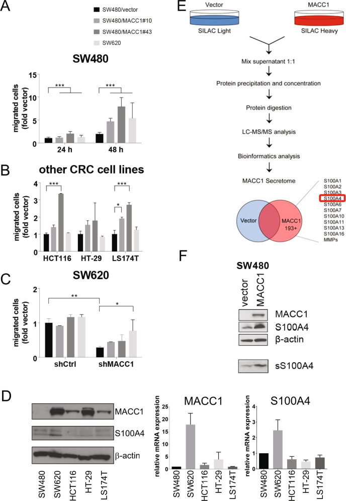
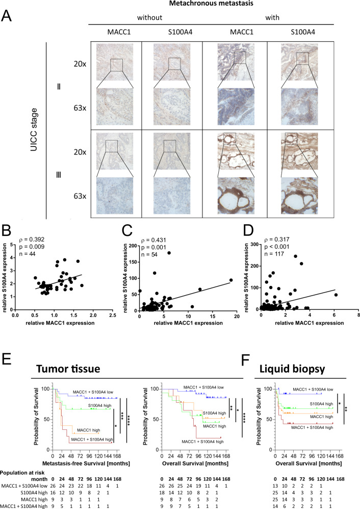
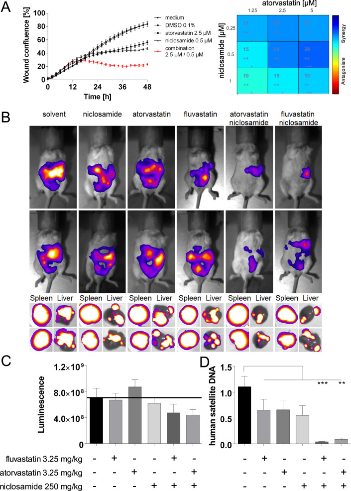
Colorectal cancer (CRC) is the second-most common malignant disease worldwide, and metastasis is the main culprit of CRC-related death. Metachronous metastases remain to be an unpredictable, unpreventable, and fatal complication, and tracing the molecular chain of events that lead to metastasis would provide mechanistically linked biomarkers for the maintenance of remission in CRC patients after curative treatment. We hypothesized, that Metastasis-associated in colorectal cancer-1 (MACC1) induces a secretory phenotype to enforce metastasis in a paracrine manner, and found, that the cell-free culture medium of MACC1-expressing CRC cells induces migration. Stable isotope labeling by amino acids in cell culture mass spectrometry (SILAC-MS) of the medium revealed, that S100A4 is significantly enriched in the MACC1-specific secretome. Remarkably, both biomarkers correlate in expression data of independent cohorts as well as within CRC tumor sections. Furthermore, combined elevated transcript levels of the metastasis genes MACC1 and S100A4 in primary tumors and in blood plasma robustly identifies CRC patients at high risk for poor metastasis-free (MFS) and overall survival (OS). Mechanistically, MACC1 strengthens the interaction of β-catenin with TCF4, thus inducing S100A4 synthesis transcriptionally, resulting in elevated secretion to enforce cell motility and metastasis. In cell motility assays, S100A4 was indispensable for MACC1-induced migration, as shown via knock-out and pharmacological inhibition of S100A4. The direct transcriptional and functional relationship of MACC1 and S100A4 was probed by combined targeting with repositioned drugs. In fact, the MACC1-β-catenin-S100A4 axis by statins (MACC1) and niclosamide (S100A4) synergized in inhibiting cancer cell motility in vitro and metastasis in vivo. The MACC1-β-catenin-S100A4 signaling axis is causal for CRC metastasis. Selectively repositioned drugs synergize in restricting MACC1/S100A4-driven metastasis with cross-entity potential.
© 2022. The Author(s).
Conflict of interest statement
All experiments were carried out in accordance with the guidelines approved by the institutional review board, number AA3/03/45, of the Charité—Universitätsmedizin Berlin, Germany. All patients gave written informed consent and the authors complied with all relevant ethical regulations for research involving human participants. For all studies, the welfare of the animals was maintained in accordance with the general principles governing the use of animals in experiments of the European Communities and German legislation. The study was performed in accordance to the United Kingdom Coordinating Committee on Cancer Research (UKCCCR) regulations for the Welfare of Animals and of the German Animal Protection Law and approved by the local responsible authorities, Berlin, Germany (State Office of Health and Social Affairs, Berlin, Germany), REG0010/19, G0333/18.CS works for NMI TT Pharmaservices, who offer DigiWest as a service. BK, HR, FB, SB, UK, MD, WW, GD, DK, and US declare no competing interests.
Figures


Similar articles
-
Cantharidin and Its Analogue Norcantharidin Inhibit Metastasis-Inducing Genes S100A4 and MACC1.Int J Mol Sci. 2023 Jan 7;24(2):1179. doi: 10.3390/ijms24021179. Int J Mol Sci. 2023. PMID: 36674695 Free PMC article.
-
Phase II trial to investigate the safety and efficacy of orally applied niclosamide in patients with metachronous or sychronous metastases of a colorectal cancer progressing after therapy: the NIKOLO trial.BMC Cancer. 2018 Mar 15;18(1):297. doi: 10.1186/s12885-018-4197-9. BMC Cancer. 2018. PMID: 29544454 Free PMC article. Clinical Trial.
-
S100A4 contributes to colorectal carcinoma aggressive behavior and to chemoradiotherapy resistance in locally advanced rectal carcinoma.Sci Rep. 2024 Dec 28;14(1):31338. doi: 10.1038/s41598-024-82814-9. Sci Rep. 2024. PMID: 39732925 Free PMC article.
-
S100A4 in Cancer Metastasis: Wnt Signaling-Driven Interventions for Metastasis Restriction.Cancers (Basel). 2016 Jun 20;8(6):59. doi: 10.3390/cancers8060059. Cancers (Basel). 2016. PMID: 27331819 Free PMC article. Review.
-
MACC1 - more than metastasis? Facts and predictions about a novel gene.J Mol Med (Berl). 2010 Jan;88(1):11-8. doi: 10.1007/s00109-009-0537-1. Epub 2009 Sep 30. J Mol Med (Berl). 2010. PMID: 19787327 Review.
Cited by
-
Epithelial-to-mesenchymal transition (EMT) and cancer metastasis: the status quo of methods and experimental models 2025.Mol Cancer. 2025 Jun 7;24(1):167. doi: 10.1186/s12943-025-02338-2. Mol Cancer. 2025. PMID: 40483504 Free PMC article. Review.
-
Parecoxib and 5-Fluorouracil Synergistically Inhibit EMT and Subsequent Metastasis in Colorectal Cancer by Targeting PI3K/Akt/NF-κB Signaling.Biomedicines. 2024 Jul 9;12(7):1526. doi: 10.3390/biomedicines12071526. Biomedicines. 2024. PMID: 39062099 Free PMC article.
-
The role of calcium signaling in organotropic metastasis of cancer.Acta Pharmacol Sin. 2025 Jul;46(7):1801-1812. doi: 10.1038/s41401-025-01537-3. Epub 2025 Mar 25. Acta Pharmacol Sin. 2025. PMID: 40133629 Review.
-
Chemoprophylaxis of precancerous lesions in patients who are at a high risk of developing colorectal cancer (Review).Med Int (Lond). 2024 Mar 27;4(3):25. doi: 10.3892/mi.2024.149. eCollection 2024 May-Jun. Med Int (Lond). 2024. PMID: 38628384 Free PMC article. Review.
-
MACC1-induced migration in tumors: Current state and perspective.Front Oncol. 2023 Mar 27;13:1165676. doi: 10.3389/fonc.2023.1165676. eCollection 2023. Front Oncol. 2023. PMID: 37051546 Free PMC article. Review.
References
-
- Siegel RL, Miller KD, Goding Sauer A, Fedewa SA, Butterly LF, Anderson JC, et al. Colorectal cancer statistics, 2020. CA Cancer J Clin. 2020;70:145–64. - PubMed
-
- Siegel RL, Miller KD, Jemal A. Cancer statistics, 2020. CA Cancer J Clin. 2020;70:7–30. - PubMed
-
- Pita-Fernández S, Alhayek-Aí M, González-Martín C, López-Calviño B, Seoane-Pillado T, Pértega-Díaz S. Intensive follow-up strategies improve outcomes in nonmetastatic colorectal cancer patients after curative surgery: a systematic review and meta-analysis. Ann Oncol J Eur Soc Med Oncol. 2015;26:644–56. - PubMed
-
- Stein U, Walther W, Arlt F, Schwabe H, Smith J, Fichtner I, et al. MACC1, a newly identified key regulator of HGF-MET signaling, predicts colon cancer metastasis. Nat Med. 2009;15:59–67. - PubMed
Publication types
MeSH terms
Substances
LinkOut - more resources
Full Text Sources
Medical
Research Materials
