

Hydrogen peroxide initiates oxidative stress and proteomic alterations in meningothelial cells
- PMID: 36008468
- PMCID: PMC9411503
- DOI: 10.1038/s41598-022-18548-3
Hydrogen peroxide initiates oxidative stress and proteomic alterations in meningothelial cells
Abstract
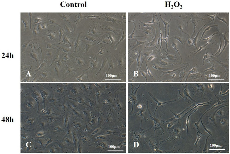
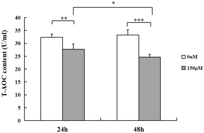
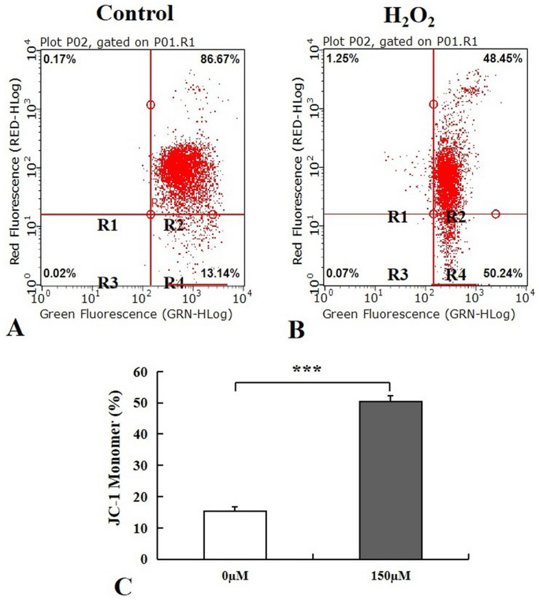
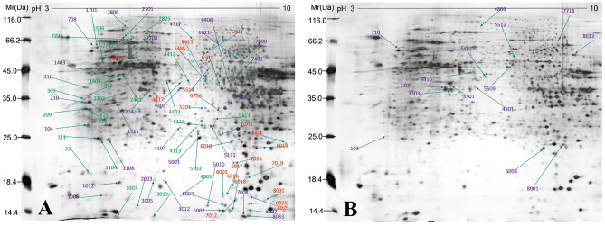
Meningothelial cells (MECs) are fundamental cells of the sheaths covering the brain and optic nerve, where they build a brain/optic nerve-cerebral spinal fluid (CSF) barrier that prevents the free flow of CSF from the subarachnoid space, but their exact roles and underlying mechanisms remain unclear. Our attempt here was to investigate the influence elicited by hydrogen peroxide (H2O2) on functional changes of MECs. Our study showed that cell viability of MECs was inhibited after cells were exposed to oxidative agents. Cells subjected to H2O2 at the concentration of 150 µM for 24 h and 48 h exhibited an elevation of reactive oxygen species (ROS) activity, decrease of total antioxidant capacity (T-AOC) level and reduced mitochondrial membrane potential (ΔΨm) compared with control cells. 95 protein spots with more than twofold difference were detected in two dimensional electrophoresis (2DE) gels through proteomics assay following H2O2 exposure for 48 h, 10 proteins were identified through TOF/MS analysis. Among the proteomic changes explored, 8 proteins related to energy metabolism, mitochondrial function, structural regulation, and cell cycle control were downregulated. Our study provides key insights that enhance our understanding of the role of MECs in the pathology of brain and optic nerve disorders.
© 2022. The Author(s).
Conflict of interest statement
The authors declare no competing interests.
Figures


References
Publication types
MeSH terms
Substances
LinkOut - more resources
Full Text Sources
