

Comment
doi: 10.1038/s42003-022-03669-2.
How many SARS-CoV-2 "viroporins" are really ion channels?
Affiliations
- PMID: 36008538
- PMCID: PMC9411608
- DOI: 10.1038/s42003-022-03669-2
Item in Clipboard
Comment
How many SARS-CoV-2 "viroporins" are really ion channels?
Commun Biol.
.
No abstract available
Conflict of interest statement
The authors declare no competing interests.
Figures
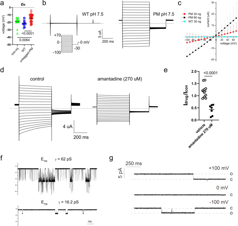
a Scatter plot of the resting (unclamped) membrane potential (EM) for Xenopus oocytes expressing untagged-PM (n = 35), WT (n = 20) or non-injected (non-inj) oocytes (triangles, n = 20) (statistical analysis by one-way ANOVA (P = 0.0002) and Tukey’s post hoc test, untagged-PM vs. WT, P < 0.0001; untagged-PM vs. non-inj, P = 0.0064; WT vs. non-inj, P = 0.3290). Data reproduced by permission from ref. . b Exemplar current traces for oocytes expressing WT (left panel) and untagged-PM (right panel) as indicated, at pH 7.5 (30 ng cRNA). Voltage protocol and scale bars are shown in the inset. Dashed lines indicate zero current level. Data reproduced by permission from ref. . c Large amplitude (µA) membrane currents on the expression of SARS-CoV-2 E protein are proportional to the quantity of RNA injected. Mean peak current versus voltage for oocytes after injection of 30 ng (red squares, n = 17) or 60 ng (black squares, n = 8) “untagged-PM” E protein cRNA, or after injection of 30 ng “WT” E protein cRNA (circles, n = 15). cRNA encoding WT and untagged-PM constructs was generated from cDNA in the pXOOM vector. Current data shown are mean ± SD, and are reproduced by permission from ref. . d Exemplar current traces for oocytes expressing untagged-PM E protein in the absence (control) or presence of amantadine (270 µM) at pH 7.5 (30 ng cRNA). Voltage protocol and scale bars are shown in the inset. Dashed lines indicate zero current level. Other experimental methods are described in ref. . e Scatter plot of fractional current at −80 mV remaining after incubation of oocytes expressing untagged-PM E protein in bath solution (vehicle) (n = 10) or amantadine (270 µM) (n = 7) as in (d). Statistical analysis performed by unpaired, two-tailed t test; bars indicate mean; error bars indicate SEM. f Single-channel currents recorded on reconstitution of a synthetic transmembrane (TM) fragment (amino acids 8–38) of E protein into artificial bilayers. The lipoprotein particles used to deliver the TM fragment into the lipid bilayer were prepared by using the protocol described in ref. for the pentameric structure formation. Recordings were made in symmetrical 300 mM NaCl, 5 mM MgCl2, 10 mM HEPES, pH 7.2, and low-pass filtered at 50 Hz for display purposes. g Single-channel currents following the reconstitution of recombinant E protein into artificial bilayers using lipid nanodiscs. E protein from cell-free protein expression in presence of lipid nanodiscs recorded in suspended lipid bilayer 4:1 DPhPC:DPhPS, in symmetrical 250 mM KCl, 10 mM HEPES, pH 7.4, 1 mM EGTA at +100 mV (upper trace), 0 mV (middle trace) and −100 mV (lower trace). Data were low-pass filtered at 500 Hz.
Comment in
-
Reply to: How Many SARS-CoV-2 "Viroporins" Are Really Ion Channels?Commun Biol. 2022 Aug 25;5(1):860. doi: 10.1038/s42003-022-03670-9. Commun Biol. 2022. PMID: 36008476 Free PMC article. No abstract available.
Comment on
-
Amantadine has potential for the treatment of COVID-19 because it inhibits known and novel ion channels encoded by SARS-CoV-2.Commun Biol. 2021 Dec 1;4(1):1347. doi: 10.1038/s42003-021-02866-9. Commun Biol. 2021. PMID: 34853399 Free PMC article.
Similar articles
-
Reply to: How Many SARS-CoV-2 "Viroporins" Are Really Ion Channels?Commun Biol. 2022 Aug 25;5(1):860. doi: 10.1038/s42003-022-03670-9. Commun Biol. 2022. PMID: 36008476 Free PMC article. No abstract available.
-
Viroporins: Structure, function, and their role in the life cycle of SARS-CoV-2.Int J Biochem Cell Biol. 2022 Apr;145:106185. doi: 10.1016/j.biocel.2022.106185. Epub 2022 Feb 24. Int J Biochem Cell Biol. 2022. PMID: 35219876 Free PMC article. Review.
-
Unravelling the Immunomodulatory Effects of Viral Ion Channels, towards the Treatment of Disease.Viruses. 2021 Oct 27;13(11):2165. doi: 10.3390/v13112165. Viruses. 2021. PMID: 34834972 Free PMC article. Review.
-
Weak Point of SARS-CoV-2: Human and Viral Ion Channels under External Physical Fields.Int J Mol Sci. 2022 Dec 2;23(23):15185. doi: 10.3390/ijms232315185. Int J Mol Sci. 2022. PMID: 36499511 Free PMC article.
-
ORF8a as a viroporin in SARS-CoV-2 infection?Cytokine Growth Factor Rev. 2021 Oct;61:1. doi: 10.1016/j.cytogfr.2021.07.002. Epub 2021 Aug 2. Cytokine Growth Factor Rev. 2021. PMID: 34362671 Free PMC article. No abstract available.
Cited by
-
Endogenous currents in HEK 293 cells are inhibited by memantine.Nat Chem Biol. 2023 Nov;19(11):1303-1305. doi: 10.1038/s41589-023-01423-1. Epub 2023 Oct 5. Nat Chem Biol. 2023. PMID: 37798356 No abstract available.
-
In silico determination of novel SARS-CoV-2 envelope protein ion channel inhibitors.Comput Struct Biotechnol J. 2025 Jun 26;27:2823-2831. doi: 10.1016/j.csbj.2025.06.036. eCollection 2025. Comput Struct Biotechnol J. 2025. PMID: 40677240 Free PMC article.
-
Anti-SARS-CoV-2 Activity of Adamantanes In Vitro and in Animal Models of Infection.COVID. 2022 Nov;2(11):1551-1563. doi: 10.3390/covid2110111. Epub 2022 Oct 28. COVID. 2022. PMID: 37274537 Free PMC article.
-
To Be or Not to Be an Ion Channel: Cryo-EM Structures Have a Say.Cells. 2023 Jul 17;12(14):1870. doi: 10.3390/cells12141870. Cells. 2023. PMID: 37508534 Free PMC article. Review.
-
Reply to: How Many SARS-CoV-2 "Viroporins" Are Really Ion Channels?Commun Biol. 2022 Aug 25;5(1):860. doi: 10.1038/s42003-022-03670-9. Commun Biol. 2022. PMID: 36008476 Free PMC article. No abstract available.
References
-
- Schulze, T. et al. SARS-CoV-2 envelope-protein corruption of homeostatic signaling mechanisms in mammalian cells. Preprint at https://www.biorxiv.org/content/10.1101/2021.06.16.448640v1 (2021). - DOI
