. 2022 Aug 25;5(1):867.
doi: 10.1038/s42003-022-03833-8.

Repeated intravenous administration of hiPSC-MSCs enhance the efficacy of cell-based therapy in tissue regeneration

Affiliations

Repeated intravenous administration of hiPSC-MSCs enhance the efficacy of cell-based therapy in tissue regeneration

Si-Jia Sun et al. Commun Biol. .

Abstract

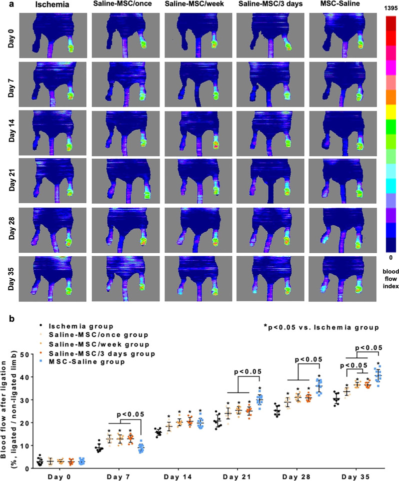
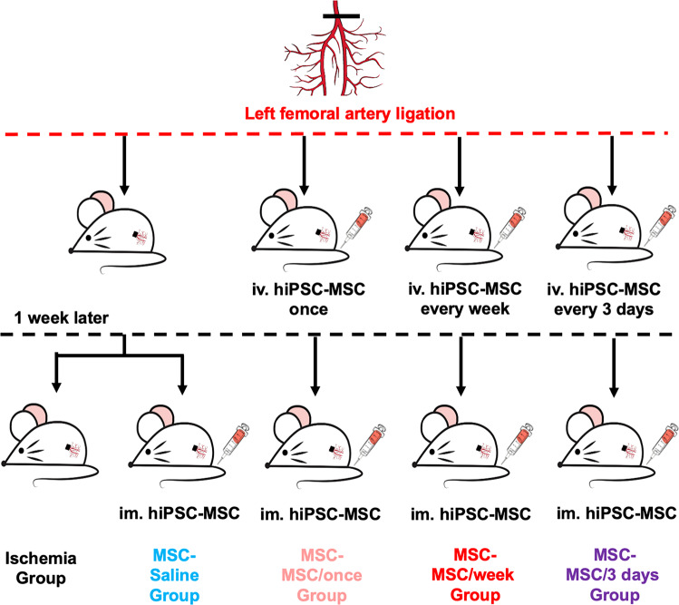
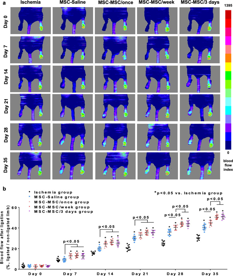
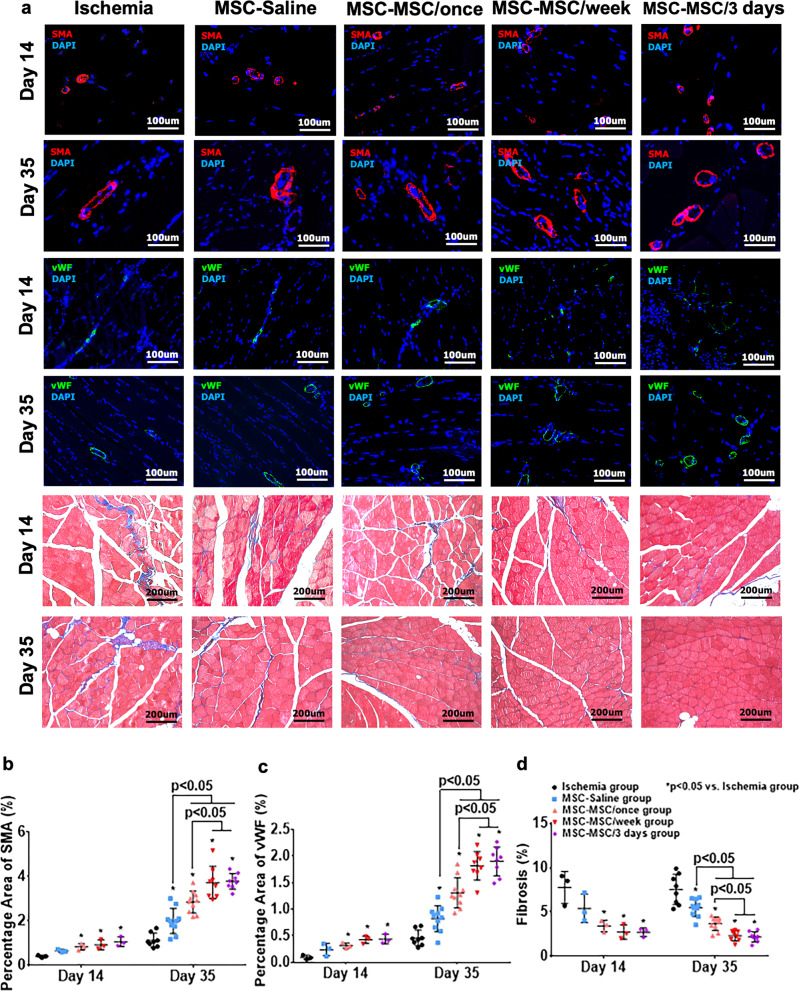
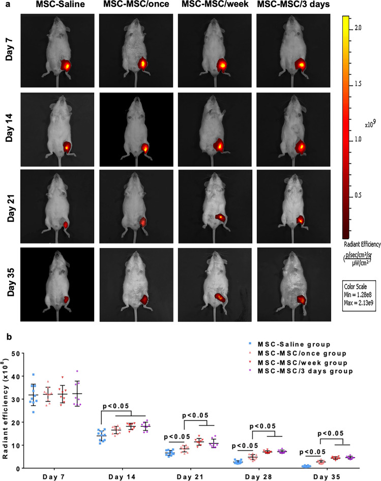
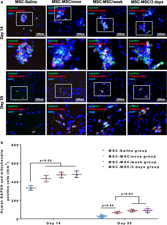
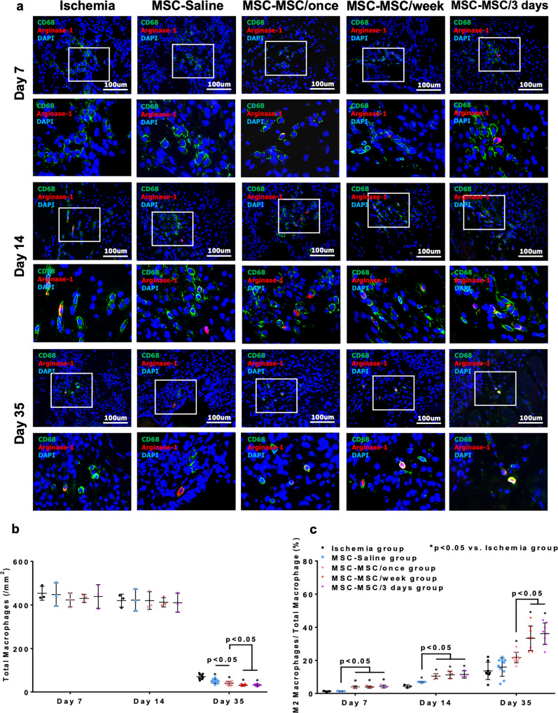
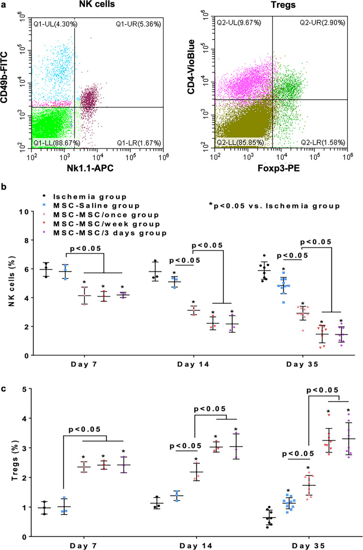
We seek to demonstrate whether therapeutic efficacy can be improved by combination of repeated intravenous administration and local transplantation of human induced pluripotential stem cell derived MSCs (hiPSC-MSCs). In this study, mice model of hind-limb ischemia is established by ligation of left femoral artery. hiPSC-MSCs (5 × 10⁵) is intravenously administrated immediately after induction of hind limb ischemia with or without following intravenous administration of hiPSC-MSCs every week or every 3 days. Intramuscular transplantation of hiPSC-MSCs (3 × 10⁶) is performed one week after induction of hind-limb ischemia. We compare the therapeutic efficacy and cell survival of intramuscular transplantation of hiPSC-MSCs with or without a single or repeated intravenous administration of hiPSC-MSCs. Repeated intravenous administration of hiPSC-MSCs can increase splenic regulatory T cells (Tregs) activation, decrease splenic natural killer (NK) cells expression, promote the polarization of M2 macrophages in the ischemic area and improved blood perfusion in the ischemic limbs. The improved therapeutic efficacy of MSC-based therapy is due to both increased engraftment of intramuscular transplanted hiPSC-MSCs and intravenous infused hiPSC-MSCs. In conclusion, our study support a combination of repeated systemic infusion and local transplantation of hiPSC-MSCs for cardiovascular disease.


Conflict of interest statement

The authors declare no competing interests.

Figures

Fig. 1
Fig. 1. Comparison of intravenous administration of hiPSC-MSCs versus intramuscular hiPSC-MSCs delivery.
To evaluate blood perfusion in the groups that received intravenous hiPSC-MSCs infusion without intramuscular hiPSC-MSCs transplantation, Laser Doppler imaging analysis was performed immediately and every week following femoral artery ligation (a). A single or repeated intravenous administration of hiPSC-MSCs in the Saline-MSC/once, Saline-MSC/week or Saline-MSC/3 days groups significantly increased blood perfusion from day 7 onwards compared with the ischemia group. Moreover, repeated intravenous hiPSC-MSCs infusion further improved blood perfusion at day 35. Nonetheless intramuscular hiPSC-MSC transplantation in the MSC-Saline group showed a superior beneficial effect over repeated intravenous hiPSC-MSC infusion in the Saline-MSC/week and Saline-MSC/3 days groups (b).
Fig. 2
Fig. 2. Flow chart of the experiment.
There are five groups of ICR mice in main experiment: ischemia group, MSC-Saline group, MSC-MSC/once group, MSC-MSC/week group, MSC-MSC/3 days group.
Fig. 3
Fig. 3. Therapeutic efficacy of intravenous cellular administration combined with intramuscular cellular delivery.
Laser Doppler imaging analysis was performed immediately and every week following femoral artery ligation to evaluate blood perfusion in the ischemic hind limbs (a). After intramuscular transplantation of hiPSC-MSCs, blood perfusion was significantly improved in the MSC-Saline, MSC-MSC/once, MSC-MSC/week and MSC-MSC/3 days groups compared with the ischemia group from day 14 onwards (all p < 0.05). A single and repeated intravenous hiPSC-MSC infusion further improved blood perfusion in the MSC-MSC/once, MSC-MSC/week and MSC-MSC/3 days groups compared with MSC-Saline group (all p < 0.05). Moreover, the blood perfusion was significantly higher in the MSC-MSC/week and MSC-MSC/3 days groups compared with the MSC-MSC/once group (all p < 0.05). There was no significant difference between the MSC-MSC/week and MSC-MSC/3 days groups (p > 0.05) (b).
Fig. 4
Fig. 4. Repeated intravenous administration of hiPSC-MSCs increased neovascularization and decreased fibrosis.
Immunohistochemical staining with anti-mouse vWF (green) and anti-mouse α-SMA (red) antibodies was performed to assess angiogenesis and arteriogenesis in ischemic tissues. Masson’s trichrome staining was performed to evaluate the degree of fibrosis (a). On day 14, neovascularization was markedly increased in the MSC-MSC/once, MSC-MSC/week and MSC-MSC/3 days groups, not in the MSC-Saline group, relative to the ischemia group. On day 35, after intramuscular transplantation of hiPSC-MSCs, neovascularization was significantly improved in the MSC-Saline, MSC-MSC/once, MSC-MSC/week and MSC-MSC/3 days groups compared with the ischemia group (all p < 0.05). Intravenous administration of hiPSC-MSCs in the MSC-MSC/once, MSC-MSC/week and MSC-MSC/3 days groups enhanced the therapeutic effects of intramuscularly transplanted hiPSC-MSCs on neovascularization compared with the MSC-Saline group (all p < 0.05). Moreover, neovascularization was further enhanced by repeated intravenous hiPSC-MSC infusion in the MSC-MSC/week and MSC-MSC/3 days groups compared with the MSC-MSC/once group (b, c). On day 14, fibrosis was remarkably decreased in the MSC-MSC/once, MSC-MSC/week and MSC-MSC/3 days groups, not in the MSC-Saline group, relative to the ischemia group. On day 35, after intramuscular transplantation of hiPSC-MSCs, fibrosis was significantly reduced in the MSC-Saline, MSC-MSC/once, MSC-MSC/week and MSC-MSC/3 days groups compared with the ischemia group (all p < 0.05). Intravenous administration of hiPSC-MSCs in the MSC-MSC/once, MSC-MSC/week and MSC-MSC/3 days groups enhanced the therapeutic effects of intramuscularly transplanted hiPSC-MSCs on reduction of fibrosis compared with the MSC-Saline group (all p < 0.05). Moreover, the anti-fibrotic effect was further enhanced by repeated intravenous hiPSC-MSC infusion in the MSC-MSC/week and MSC-MSC/3 days groups compared with the MSC-MSC/once group (d).
Fig. 5
Fig. 5. Repeated intravenous administration of hiPSC-MSCs prolonged the survival of transplanted hiPSC-MSCs.
A series of fluorescent images of ischemic hind limbs was performed immediately and every week following intramuscular transplantation of hiPSC-MSCs to detect the fate of intramuscularly transplanted hiPSC-MSCs (a). A single or repeated intravenous hiPSC-MSCs infusion in the MSC-MSC/once, MSC-MSC/week and MSC-MSC/3 days groups significantly prolonged the survival of intramuscular transplanted hiPSC-MSCs from day 14 onwards compared with the MSC-Saline group (all p < 0.05). Moreover, repeated intravenous hiPSC-MSCs infusion in the MSC-MSC/week and MSC-MSC/3 days groups further improved the survival of intramuscularly transplanted hiPSC-MSCs from day 21 onwards compared with the MSC-MSC/once group (all p < 0.05), whereas no significant difference was observed between MSC-MSC/week and MSC-MSC/3 days groups (p > 0.05) (b).
Fig. 6
Fig. 6. Repeated intravenous administration of hiPSC-MSCs improved the engraftment of transplanted hiPSC-MSCs in ischemic tissue.
The engraftment of intramuscularly transplanted hiPSC-MSCs was further confirmed by double immunohistochemical staining with anti-human GAPDH (green) and anti-human mitochondria antibodies (red) at day 14 and 35 (a). A single or repeated intravenous hiPSC-MSC infusion in the MSC-MSC/once, MSC-MSC/week and MSC-MSC/3 days groups significantly improved the engraftment of intramuscularly transplanted hiPSC-MSCs from day 14 onwards (all p < 0.05). Repeated intravenous hiPSC-MSC infusion in the MSC-MSC/week and MSC-MSC/3 days groups further improved the engraftment of intramuscular transplanted hiPSC-MSCs at day 35 compared with the MSC-MSC/once group (all p < 0.05), whereas no significant difference was observed between the MSC-MSC/week and MSC-MSC/3 days groups (p > 0.05) (b).
Fig. 7
Fig. 7. Repeated intravenous administration of hiPSC-MSCs reduced the infiltration of macrophages and promoted polarization of M2 macrophages.
Muscular infiltration of macrophages was determined by immunohistochemical staining with anti-mouse CD68 antibody (green) at day 7, 14, and 35. Number of M2 macrophages was detected by immunohistochemical staining with anti-mouse Arginase-1 antibodies (red) (a). At day 35, after intramuscular transplantation of hiPSC-MSCs, total macrophages were significantly decreased in the MSC-Saline, MSC-MSC/once, MSC-MSC/week and MSC-MSC/3 days groups compared with the ischemia group (all p < 0.05). A single or repeated intravenous hiPSC-MSCs infusion in the MSC-MSC/once, MSC-MSC/week and MSC-MSC/3 days groups significantly decreased the muscular infiltration of macrophages compared with the MSC-Saline group (all p < 0.05). In addition, repeated intravenous hiPSC-MSCs infusion in the MSC-MSC/week and MSC-MSC/3 days groups further decreased the muscular infiltration of macrophages compared with the MSC-MSC/once group (all p < 0.05). Nevertheless no significant difference was observed between groups at day 7 and 14 (all p > 0.05) (b). Intramuscular transplantation of hiPSC-MSCs without intravenous hiPSC-MSC infusion significantly increased the polarization of M2 macrophages at day 14 compared with the ischemia group (p < 0.05). A single or repeated intravenous hiPSC-MSC infusion in the MSC-MSC/once, MSC-MSC/week and MSC-MSC/3 days groups significantly improved the polarization of M2 macrophages from day 7 onwards (all p < 0.05). Repeated hiPSC-MSCs infusion further promoted the polarization of M2 macrophages compared with a single intravenous hiPSC-MSCs infusion in the MSC-MSC/once group at day 35 (all p < 0.05) (c).
Fig. 8
Fig. 8. Repeated intravenous administration of hiPSC-MSCs reduced splenic NK cells and increased splenic Tregs.
Splenic Tregs and NK cells were determined by flow cytometry analysis at day 7, 14 and 35 (a). After intramuscular transplantation of hiPSC-MSCs, splenic NK cells were significantly decreased in the MSC-Saline, MSC-MSC/once, MSC-MSC/week and MSC-MSC/3 days groups from day 14 onwards compared with the ischemia group (all p < 0.05). A single or repeated intravenous hiPSC-MSC infusion in the MSC-MSC/once, MSC-MSC/week and MSC-MSC/3 days groups significantly decreased splenic NK cells from day 7 onwards compared with the ischemia and MSC-Saline groups (all p < 0.05). Repeated intravenous hiPSC-MSC infusion in the MSC-MSC/week and MSC-MSC/3 days groups further decreased splenic NK cells from day 14 onwards compared with the MSC-MSC/once group (all p < 0.05) (b). After intramuscular transplantation of hiPSC-MSCs, splenic Tregs were significantly increased in the MSC-Saline, MSC-MSC/once, MSC-MSC/week and MSC-MSC/3 days groups at day 35 compared with the ischemia group (all p < 0.05). A single or repeated intravenous hiPSC-MSC infusion in the MSC-MSC/once, MSC-MSC/week and MSC-MSC/3 days groups significantly increased splenic Tregs compared with the ischemia and MSC-Saline groups (all p < 0.05). Moreover, repeated intravenous hiPSC-MSC infusion in the MSC-MSC/week and MSC-MSC/3 days groups further increased splenic Tregs from day 14 onwards compared with the MSC-MSC/once group (all p < 0.05) (c).

Similar articles

Cited by

References

    1. Menasche P. Cell therapy trials for heart regeneration - lessons learned and future directions. Nat. Rev. Cardiol. 2018;15:659–671. doi: 10.1038/s41569-018-0013-0. - DOI - PubMed
    1. Kim S-W, et al. Cultured Human Bone Marrow-Derived CD31+ Cells Are Effective for Cardiac and Vascular Repair Through Enhanced Angiogenic, Adhesion, and Anti-Inflammatory Effects. J. Am. Coll. Cardiol. 2014;64:1681–1694. doi: 10.1016/j.jacc.2014.06.1204. - DOI - PMC - PubMed
    1. Grigorian Shamagian L, et al. Perspectives on Directions and Priorities for Future Preclinical Studies in Regenerative Medicine. Circ. Res. 2019;124:938–951. doi: 10.1161/CIRCRESAHA.118.313795. - DOI - PMC - PubMed
    1. Vazir A, et al. Can we remove scar and fibrosis from adult human myocardium? Eur. Heart J. 2019;40:960–966. doi: 10.1093/eurheartj/ehy503. - DOI - PubMed
    1. Madonna R, et al. Position Paper of the European Society of Cardiology Working Group Cellular Biology of the Heart: Cell-based therapies for myocardial repair and regeneration in ischemic heart disease and heart failure. Eur. Heart J. 2016;37:1789–1798. doi: 10.1093/eurheartj/ehw113. - DOI - PMC - PubMed

Publication types