. 2022 Aug 26;41(1):259.
doi: 10.1186/s13046-022-02471-6.

Irradiation induces DJ-1 secretion from esophageal squamous cell carcinoma cells to accelerate metastasis of bystander cells via a TGF-β1 positive feedback loop

Affiliations

Irradiation induces DJ-1 secretion from esophageal squamous cell carcinoma cells to accelerate metastasis of bystander cells via a TGF-β1 positive feedback loop

Junjie Gu et al. J Exp Clin Cancer Res. .

Abstract

Background: Radiation-induced bystander effect (RIBE) can promote tumor metastasis contributing to the failure of radiotherapy for esophageal squamous cell carcinoma (ESCC). Aberrant expression of DJ-1 has been identified in ESCC; however, the relationship between DJ-1 and RIBE in ESCC remains unknown.

Methods: We detected DJ-1 in the serum and cell supernatants by enzyme-linked immunosorbent assay (ELISA) and evaluated tumor metastasis by phenotypic experiments in vivo and in vitro. RNA-seq, mass spectrometry, western blot (WB), immunoprecipitation (IP), and dual-luciferase reporter assays were performed to explore the underlying mechanisms.

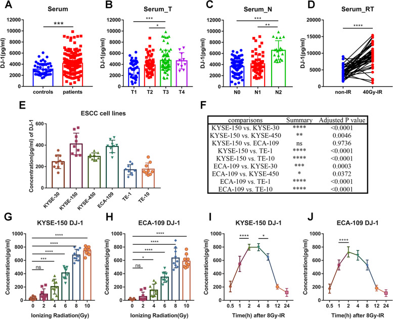
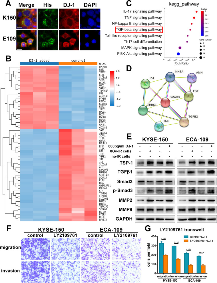
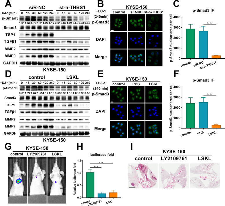
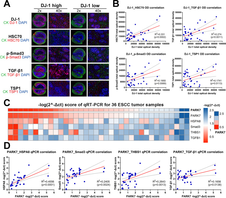
Results: DJ-1 was highly expressed in the serum of patients with ESCC receiving radiotherapy and was significantly overexpressed in the medium of ESCC cells receiving irradiation. DJ-1 promoted tumor metastasis via the TGF-β1 pathway. Mechanistic studies revealed that DJ-1 bound to HSC70 to promote Smad3 phosphorylation and nuclear aggregation in a protein-interaction manner, which activated the transcription of Thrombospondin-1 (TSP1). Subsequently, the activation of TGF-β1 by TSP1 re-promoted Smad3 phosphorylation and nuclear aggregation, constituting a positive feedback loop to strengthen the metastasis of ESCC cells, which was effectively blocked by LY2109761 and LSKL. Moreover, higher levels of serum DJ-1 in patients with ESCC were related to a poorer prognosis of radiotherapy.

Conclusions: Irradiation can induce ESCC cells secreting DJ-1. Secreted DJ-1 enters bystander cells to initiate activation of the TGF-β1 pathway via the DJ-1/HSC70/Smad3 signaling axis. The TSP1/TGF-β1/Smad3 positive feedback pathway constitutes the core pathway that promotes ESCC metastasis. DJ-1 is a useful biomarker for predicting the efficacy of radiotherapy and a potential therapeutic target for reversing RIBE in ESCC. Schematic diagram showing the underlying mechanism that irradiation-induced secretion of DJ-1 accelerates the metastasis of bystander ESCC cells.

Keywords: DJ-1; Esophageal squamous cell carcinoma; HSC70; Radiation-induced bystander effect; Smad3; TGF-β1; TSP1; Tumor metastasis.


Conflict of interest statement

The authors declare that they have no competing interests.

Figures

Fig. 1
Fig. 1
Serum DJ-1 expression correlates tumor progression and upregulates from ESCC cells for irradiation. A ELISA results of DJ-1 levels in serum of ESCC patients (n = 177) and normal donors (n = 50). B-C The expression of DJ-1 in serum of ESCC patients at multiple stages (T and N) by ELISA. D ELISA results of DJ-1 levels in serum of ESCC patients (n = 46) at the point of before and after radiotherapy (40 Gy). E–F ELISA results of DJ-1 expression in medium of 6 ESCC cell lines at the point of 2 h after 6 Gy-irradiation. G-H ELISA results of DJ-1 expression in medium of K150 and E109 cells at the point of 2 h after receiving multiple doses (0, 2, 4, 6, 8 and 10 Gy) of irradiation. I-J ELISA results of DJ-1 expression in medium of K150 and E109 cells at multiple timing (0.5, 1, 2, 4, 8, 12 and 24 h) after receiving 8 Gy irradiation. *p < 0.05, **p < 0.01, ***p < 0.001, ****p < 0.0001
Fig. 2
Fig. 2
Exogenous DJ-1 enhances the metastasis of ESCC cells in vitro and vivo. A Representative images of transwell assays for bystander ESCC cells co-culture with irradiated cells or exogenous DJ-1. Scale bars, 100 μm. B Representative images of wound-healing assays for bystander ESCC cells co-culture with irradiated cells or exogenous DJ-1. Scale bars, 200 μm. C-D The statistical graphs of transwell assays presented in part A. E–F The statistical graphs of Wound-healing assays presented in part B. G-H Representative IF images and the statistical graph of the 3D tumor spheroid invasion assay for bystander cells (green) co-culture with irradiated cells or exogenous DJ-1. Scale bars, 200 μm. I-J Representative photographs of metastatic nodules were taken from nude mice injected DJ-1 protein by an IVIS imaging system. Quantification of the luciferase was shown. K H&E staining was used to characterize the lung metastatic nodules. Scale bar, 2 mm. **p < 0.01, ***p < 0.001
Fig. 3
Fig. 3
Exogenous DJ-1 promotes metastasis of bystander ESCC cells by activating TGF-β1 pathway. A Representative IF images of the intracellular localization of exogenous DJ-1 (red) tagged by His-tag (green) in ESCC cells detected using immunofluorescence microscopy. Scale bar, 20 μm. B Heatmaps of 59 representative DEGs screened from RNAseq analysis. C KEGG pathway enrichment analyses showed top activated gene sets ordered by rich ratio and the TGF-β1 signaling pathway was enriched after DJ-1 stimulation. D A network of proteins enriched in the TGF-β1 signaling pathway and encoded by DEGs from RNAseq results. E Western blot results of prominent proteins correlated with the TGF-β1 signaling pathway in bystander ESCC cells co-cultured with irradiated cells or DJ-1. F-G Representative images and the statistical graph of transwell assays for bystander ESCC cells measured by LY2109761. Scale bars, 100 μm. ****p < 0.0001
Fig. 4
Fig. 4
Exogenous DJ-1 promotes metastasis by binding to HSC70 in ESCC cells. A Results of proteins identified from LC–MS/MS ordered by the score of iBAQ. B The targets of intersection between proteins identified from LC–MS/MS and DEGs screened from RNAseq. C Representative IF images of colocalization of DJ-1 (red) and HSC70 (green) in K150 and E109 cells detected using immunofluorescence microscopy. Scale bar, 20 μm. D Coimmunoprecipitation analysis demonstrated that exogenous DJ-1 could interact with HSC70. E–H Representative images and the statistical graph of transwell assays for bystander K150 and E109 cells transfected with siRNA targeting HSC70. Scale bars, 100 μm. ***p < 0.001
Fig. 5
Fig. 5
Exogenous DJ-1 acting on HSC70 promotes Smad3 phosphorylation and nuclear aggregation in ESCC cells. A Western blot results of Smad3 phosphorylation status relative to total Smad3 in K150 and E109 cells transfected with siRNA targeting HSC70 at the timing (0, 15, 30, 60, 120 and 240 min) after DJ-1 stimulation. B Representative IF images of p-Smad3 (green) and HSC70 (red) expression in K150 and E109 cells transfected with siRNA targeting HSC70 at the timing (0, 15 and 30 min) after DJ-1 stimulation. Scale bar, 20 μm. C Coimmunoprecipitation analysis demonstrated that HSC70 could interact with Smad3 in attendance of DJ-1. D-E Representative images and the statistical graph of transwell assays for bystander K150 and E109 cells transfected with siRNA targeting Smad3. Scale bars, 100 μm. **p < 0.01, ***p < 0.001, ****p < 0.0001
Fig. 6
Fig. 6
Exogenous TGF-β1 reactivates Smad3 without the presence of HSC70. A-B Representative images and the statistical graph of transwell assays for bystander HSC70-knockdown K150 and E109 cells stimulated by TGF-β1. Scale bars, 100 μm. C-D Representative IF images and the statistical graph of 3D tumor spheroid invasion assays for bystander HSC70-knockdown K150 and E109 cells (green) stimulated by TGF-β1. Scale bars, 200 μm. E–F Representative IF images of p-Smad3 (green) expression in HSC70-knockdown ESCC cells stimulated by TGF-β1 at the timing (240 min) after DJ-1 stimulation. Scale bar, 20 μm. G. Western blot results of Smad3 phosphorylation status relative to total Smad3 in HSC70-knockdown ESCC cells stimulated by TGF-β1. ***p < 0.001
Fig. 7
Fig. 7
Smad3 transcribes THBS1 to promote the metastasis of K150 cells. A Western blot results of prominent proteins correlated with the TGF-β1 signaling pathway in bystander K150 cells at the timing (0, 15, 30, 60, 120 and 240 min) after DJ-1 stimulation. B Schematic representation of SMAD3-binding sites on THBS1 promoter. The binding site is located at -1956 ~ -1947 on the THBS1 promoter. C-D Luciferase promoter activity was measured by transfecting a PGL3 vector containing the full-length THBS1 promoter (Full) or inserts deleted either in binding site. K150 cells were induced by exogenous DJ-1 (C) or transfected with Smad3 overexpression plasmid (D). E Western blot results of TSP1 and Smad3 in K150 cells transfected with Smad3-knockdown siRNA. F-G Representative images and the statistical graph of transwell assays for bystander K150 cells transfected with siRNA targeting THBS1. Scale bars, 100 μm. H-I Representative images and the statistical graph of transwell assays for bystander K150 cells measured by LSKL. Scale bars, 100 μm. **p < 0.01, ***p < 0.001, ****p < 0.0001
Fig. 8
Fig. 8
Blocking TSP1 reduces phosphorylation and nuclear aggregation of Smad3 and metastasis in vivo. A Western blot results of prominent proteins correlated with the TGF-β1 signaling pathway in bystander K150 cells transfected with siRNA targeting THBS1 at the timing (0, 15, 30, 60, 120 and 240 min) after DJ-1 stimulation. B-C Representative IF images of p-Smad3 (green) expression in K150 cells transfected with siRNA targeting THBS1 at the timing (240 min) after DJ-1 stimulation. Scale bar, 20 μm. D Western blot results of prominent proteins correlated with the TGF-β1 signaling pathway in bystander K150 cells measured by LSKL at the timing (0, 15, 30, 60, 120 and 240 min) after DJ-1 stimulation. E–F Representative IF images of p-Smad3 (green) expression in K150 cells measured by LSKL at the timing (240 min) after DJ-1 stimulation. Scale bar, 20 μm. G-H Representative photographs of metastatic nodules were taken from nude mice injected LY2109761 or LSKL by an IVIS imaging system. Quantification of the luciferase was shown. I H&E staining was used to characterize the lung metastatic nodules. Scale bar, 2 mm. ***p < 0.001, ****p < 0.0001
Fig. 9
Fig. 9
DJ-1 regulatory pathway proteins correlates in patient samples. A IF staining for DJ-1, HSC70, p-Smad3, TGF-β1 and TSP1 in ESCC tissues ordered by two groups (DJ-1 high and DJ-1 low). Scale bar, 500 μm (2x) and 20 μm (40x). B Correlation analysis of integrated optical density (IOD) between the detected targets with DJ-1. C Heatmaps of the score of -log(2^-Δct) from qRT-PCR results of DJ-1, HSC70, p-Smad3, TGF-β1 and TSP1 in ESCC tissues. D Correlation analysis of the score of -log(2^-Δct) between the detected targets with DJ-1

Similar articles

Cited by

References

    1. Bray F, Ferlay J, Soerjomataram I, Siegel RL, Torre LA, Jemal A. Global cancer statistics 2018: GLOBOCAN estimates of incidence and mortality worldwide for 36 cancers in 185 countries. CA Cancer J Clin. 2018;68:394–424. doi: 10.3322/caac.21492. - DOI - PubMed
    1. Kim E, Koroukian S, Thomas CR., Jr Conditional Survival of Esophageal Cancer: An Analysis from the SEER Registry (1988–2011) J Thorac Oncol. 2015;10:1490–1497. doi: 10.1097/JTO.0000000000000649. - DOI - PMC - PubMed
    1. Kamochi N, Nakashima M, Aoki S, Uchihashi K, Sugihara H, Toda S, Kudo S. Irradiated fibroblast-induced bystander effects on invasive growth of squamous cell carcinoma under cancer-stromal cell interaction. Cancer Sci. 2008;99:2417–2427. doi: 10.1111/j.1349-7006.2008.00978.x. - DOI - PMC - PubMed
    1. Al-Abedi R, Tuncay Cagatay S, Mayah A, Brooks SA, Kadhim M. Ionising Radiation Promotes Invasive Potential of Breast Cancer Cells: The Role of Exosomes in the Process. Int J Mol Sci. 2021;22:11570. doi: 10.3390/ijms222111570. - DOI - PMC - PubMed
    1. Leu JD, Wang CY, Lo CC, Lin MY, Chang CY, Hung WC, Lin ST, Wang BS, Lee YJ. Involvement of c-Myc in low dose radiation-induced senescence enhanced migration and invasion of unirradiated cancer cells. Aging (Albany NY) 2021;13:22208–22231. doi: 10.18632/aging.203527. - DOI - PMC - PubMed

Substances