In vivo hamster cheek pouch subepithelial ablation, biomaterial injection, and localization: pilot study
- PMID: 36008882
- PMCID: PMC9407625
- DOI: 10.1117/1.JBO.27.8.080501
In vivo hamster cheek pouch subepithelial ablation, biomaterial injection, and localization: pilot study
Abstract
Significance: The creation of subepithelial voids within scarred vocal folds via ultrafast laser ablation may help in localization of injectable biomaterials toward a clinically viable therapy for vocal fold scarring.
Aim: We aim to prove that subepithelial voids can be created in a live animal model and that the ablation process does not engender additional scar formation. We demonstrate localization and long-term retention of an injectable biomaterial within subepithelial voids.
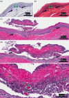
Approach: A benchtop nonlinear microscope was used to create subepithelial voids within healthy and scarred cheek pouches of four Syrian hamsters. A model biomaterial, polyethylene glycol tagged with rhodamine dye, was then injected into these voids using a custom injection setup. Follow-up imaging studies at 1- and 2-week time points were performed using the same benchtop nonlinear microscope. Subsequent histology assessed void morphology and biomaterial retention.
Results: Focused ultrashort pulses can be used to create large subepithelial voids in vivo. Our analysis suggests that the ablation process does not introduce any scar formation. Moreover, these studies indicate localization, and, more importantly, long-term retention of the model biomaterial injected into these voids. Both nonlinear microscopy and histological examination indicate the presence of biomaterial-filled voids in healthy and scarred cheek pouches 2 weeks postoperation.
Conclusions: We successfully demonstrated subepithelial void formation, biomaterial injection, and biomaterial retention in a live animal model. This pilot study is an important step toward clinical acceptance of a new type of therapy for vocal fold scarring. Future long-term studies on large animals will utilize a miniaturized surgical probe to further assess the clinical viability of such a therapy.
Keywords: ablation of tissue; hamster cheek pouch; in vivo; nonlinear microscopy; ultrafast lasers; vocal fold scarring.
Figures


Similar articles
-
Ultrafast laser surgery probe for sub-surface ablation to enable biomaterial injection in vocal folds.Sci Rep. 2022 Nov 29;12(1):20554. doi: 10.1038/s41598-022-24446-5. Sci Rep. 2022. PMID: 36446830 Free PMC article.
-
Ultrafast Laser Microlaryngeal Surgery for In Vivo Subepithelial Void Creation in Canine Vocal Folds.Laryngoscope. 2023 Nov;133(11):3042-3048. doi: 10.1002/lary.30713. Epub 2023 Apr 25. Laryngoscope. 2023. PMID: 37096749 Free PMC article.
-
Quantitative differentiation of normal and scarred tissues using second-harmonic generation microscopy.Scanning. 2016 Nov;38(6):684-693. doi: 10.1002/sca.21316. Epub 2016 Apr 25. Scanning. 2016. PMID: 27111090 Free PMC article.
-
Perspectives on adipose-derived stem/stromal cells as potential treatment for scarred vocal folds: opportunity and challenges.Curr Stem Cell Res Ther. 2010 Jun;5(2):175-81. doi: 10.2174/157488810791268591. Curr Stem Cell Res Ther. 2010. PMID: 19941448 Review.
-
Pulse dye and other laser treatments for vocal scar.Curr Opin Otolaryngol Head Neck Surg. 2010 Dec;18(6):492-7. doi: 10.1097/MOO.0b013e32833f890d. Curr Opin Otolaryngol Head Neck Surg. 2010. PMID: 20842035 Review.
Cited by
-
Ultrafast laser surgery probe for sub-surface ablation to enable biomaterial injection in vocal folds.Sci Rep. 2022 Nov 29;12(1):20554. doi: 10.1038/s41598-022-24446-5. Sci Rep. 2022. PMID: 36446830 Free PMC article.
-
Ultrafast Laser Microlaryngeal Surgery for In Vivo Subepithelial Void Creation in Canine Vocal Folds.Laryngoscope. 2023 Nov;133(11):3042-3048. doi: 10.1002/lary.30713. Epub 2023 Apr 25. Laryngoscope. 2023. PMID: 37096749 Free PMC article.
References
Publication types
MeSH terms
Substances
Grants and funding
LinkOut - more resources
Full Text Sources
Medical

