Review
. 2022 Jul 30;12(8):1060.
doi: 10.3390/biom12081060.

Nanoscale Technologies in the Fight against COVID-19: From Innovative Nanomaterials to Computer-Aided Discovery of Potential Antiviral Plant-Derived Drugs

Affiliations
Review

Nanoscale Technologies in the Fight against COVID-19: From Innovative Nanomaterials to Computer-Aided Discovery of Potential Antiviral Plant-Derived Drugs

Nunzio Iraci et al. Biomolecules. .

Abstract

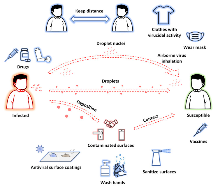
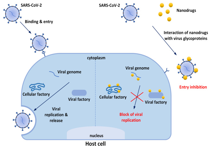
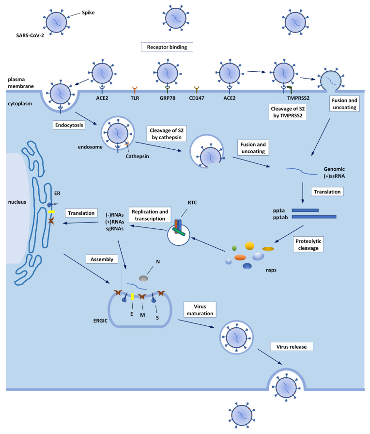
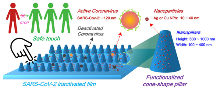
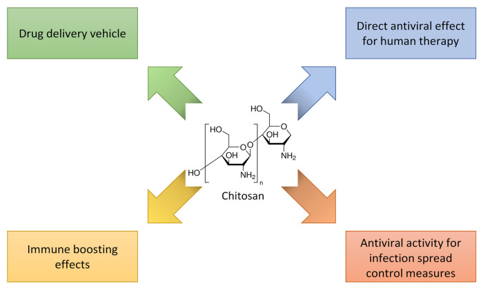
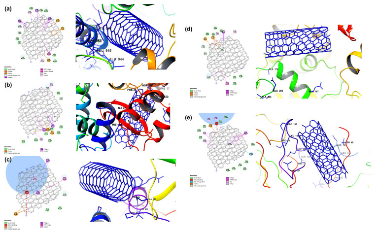
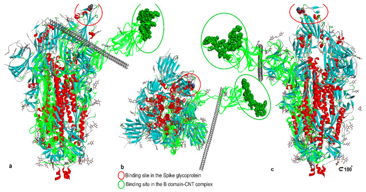
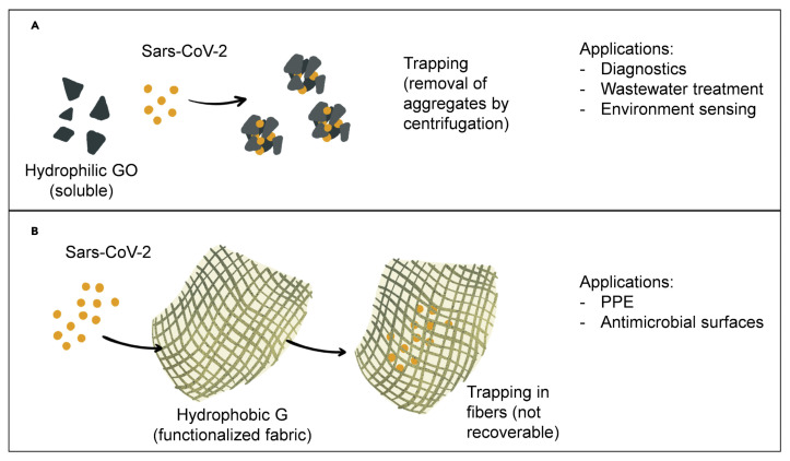
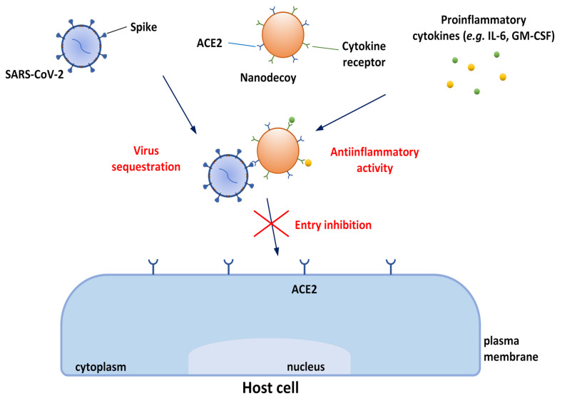
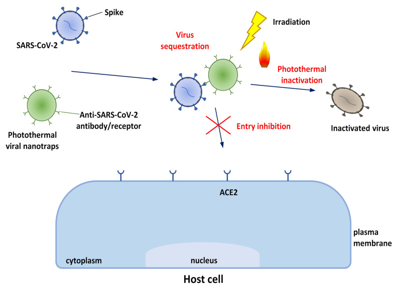
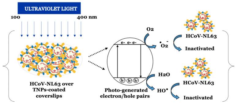
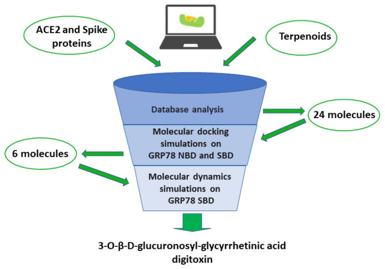
The last few years have increasingly emphasized the need to develop new active antiviral products obtained from artificial synthesis processes using nanomaterials, but also derived from natural matrices. At the same time, advanced computational approaches have found themselves fundamental in the repurposing of active therapeutics or for reducing the very long developing phases of new drugs discovery, which represents a real limitation, especially in the case of pandemics. The first part of the review is focused on the most innovative nanomaterials promising both in the field of therapeutic agents, as well as measures to control virus spread (i.e., innovative antiviral textiles). The second part of the review aims to show how computer-aided technologies can allow us to identify, in a rapid and therefore constantly updated way, plant-derived molecules (i.e., those included in terpenoids) potentially able to efficiently interact with SARS-CoV-2 cell penetration pathways.

Keywords: SARS-CoV-2; antiviral activity; drug delivery systems; molecular docking; nanodecoys; nanosystems; terpenoids; virus spread control measures.


Conflict of interest statement

The authors declare no conflict of interest.

Figures

Figure 1
Figure 1
Transmission methods of SARS-CoV-2 and possible approaches to prevent and treat COVID-19.
Figure 2
Figure 2
Possible mechanisms involved in the effectiveness of nanodrugs for prevention and treatment of COVID-19.
Figure 3
Figure 3
(a) Cryo-electron microscopy structure of SARS-CoV-2 spike protein (residues 1-1147—PDB ID: 7DCC). Spike protein domains are color coded as follows: S1—cyan; RBD—magenta; S2—yellow; FP—green; HR1—blue [53]. (b) X-ray crystal structure of ACE2 bound to SARS-CoV-2 spike protein (PDB ID: 6M0J). ACE2 is depicted in green cartoons, with the spike/ACE2 interface residues highlighted in magenta and the zinc atom represented by a blue sphere. Spike protein RBD is depicted in gray [54].
Figure 4
Figure 4
SARS-CoV-2 life cycle. SARS-CoV-2 entry into the host cell is mediated by the binding of the Spike protein (S) to ACE2. Then, entry requires S protein priming by the TMPRSS2, leading to S protein cleavage, which facilitates viral fusion with the membrane of the target cell. In case of low levels of TMPRSS2 on the target cell, the virus is internalized via endocytosis and, after endosomal acidification, S2 is cleaved by the enzymes cathepsins. Then, regardless of which of the two mechanisms was followed, the viral RNA is released into the cytoplasm of the host cell. Emerging evidence supports interactions between S protein and other host receptors and proteins (such as GRP78, CD147, TLRs), apart from ACE2, that may represent alternative routes for viral entry. After the release of viral (+)ssRNA genome into the cytoplasm, pp1a and pp1ab are produced by cellular ribosomes and then processed by viral proteases (PLpro and 3CLpro) into 16 nsps that form the replicase–transcriptase complex (RTC). The RTC mediates the synthesis of (-)RNA. A full-length (-)RNA copy serves as a template for the full-length (+)ssRNA genome, whereas sgRNAs are translated into structural (S, M, E, N) and accessory proteins. The viral nucleocapsid is assembled from newly synthesized viral genomic (+)ssRNA and N proteins in the cytoplasm, and then buds into the ER–Golgi intermediate cavity (ERGIC) reaching the S, E, and M for viral assembly. The new virions are then released from the cells via exocytosis.
Figure 5
Figure 5
Simulation outcomes concerning the structures of some RBD complexes with Au NPs. Amin: 8-mercaptooctan-1-aminium; EG2: 2-(2-(6-mercaptohexyl)oxy)ethoxy)ethan-1-ol; Mes: 3-mercaptoethylsulfonate; Mus: Undecanesulfonic acid; Mus-Ot: Undecanesulfonic acid/Octanethiol; Ot: Octanethiol; Pep: Cys-Gln-Thr-Asp-Lys-His-Glu-Glu-Asp-Tyr-Gln-Met-Lys-Gly-Asp-Arg. Reprinted with permission from A. Mehranfar and M. Izadyar. Theoretical Design of Functionalized Gold Nanoparticles as Antiviral Agents against Severe Acute Respiratory Syndrome Coronavirus 2 (SARS-CoV-2). Journal of Physical Chemistry, Letters 2020, 11, 24, 10284–10289 [98]. Copyright 2020 American Chemical Society.
Figure 6
Figure 6
Scheme of the molecular docking involving the conical Cu NPs and the target spike glycoprotein (PDB ID: 6ZGG), together with the pharmacophore. Reprinted from Journal of Molecular Structure, Vol 1253, Aallaei et al. Investigation of Cu metal nanoparticles with different morphologies to inhibit SARS-CoV-2 main protease and spike glycoprotein using Molecular Docking and Dynamics Simulation, Art. N. 132301, Copyright (2022) [102], with permission from Elsevier.
Figure 7
Figure 7
(a) Docking results of ZnO NPs interacting with amino acids of COVID-19 RdRp and (b) corresponding zoom. (c) The related ligand–protein interaction diagrams. The dashed green lines indicate hydrogen bonds. Figure reprinted from Ref. [121] under the terms of the Attribution-NonCommercial-NoDerivatives 4.0 International (CC BY-NC-ND 4.0) license.
Figure 8
Figure 8
(A) Schematic representation showing how ZnO petals added to nanoceutical cotton fabric may induce a denaturation of the spike protein; (B) The five steps for growing ZnO nanoflowers on cotton cellulose fibers. Reprinted with permission from Adhikari et al. Nanoceutical Fabric Prevents COVID-19 Spread through Expelled Respiratory Droplets: A Combined Computational, Spectroscopic, and Antimicrobial Study. ACS Applied Bio Materials, 2021 4 (7), 5471–5484. Copyright 2021 American Chemical Society [122].
Figure 9
Figure 9
Simplified scheme about the design of functionalized nanoscale conical pillars with antiviral properties. Figure reused from Ref. [134] under the terms of CC BY 4.0 license.
Figure 10
Figure 10
Possible applications of chitosan in COVID-19.
Figure 11
Figure 11
Molecular docking outcomes concerning the interaction potential of carbon nanotubes towards the following targets of SARS-CoV-2: (a) spike glycoprotein, (b) RdRp, (c) Mpro, (d) papain-like protease and (e) RNA-binding domain of nucleocapsid protein. Figure reprinted from Ref. [168] under the terms of the Creative Commons CC-BY-NC-ND license.
Figure 12
Figure 12
(ac) MD simulation outcomes showing the evolution of the interactions between SWCNTs and SARS-CoV-2 spike glycoprotein. The green chain corresponds to the B domain of the spike glycoprotein simulated in the presence of SWCNTs, whereas the blue–red one is the spike glycoprotein (PDB ID: 6VYB). Reprinted from Computers in Biology and Medicine, Vol 136, Jomhori et al. Tracking the interaction between single-wall carbon nanotube and SARS-Cov-2 spike glycoprotein: A molecular dynamics simulations study, Art. N. 104692, Copyright (2021) [169], with permission from Elsevier.
Figure 13
Figure 13
Schematic preparation and potentential applications of nanohybrid systems based on (A) hydrophilic GO and (B) hydrophobic graphene materials employed to defeat SARS-CoV-2. Figure reprinted from Ref. [173] under the terms of the Creative Commons CC-BY-NC-ND license.
Figure 14
Figure 14
Cytomimetic nanosystems are innovative materials in the fight against COVID-19. The present figure is a schematic representation of the nanodecoy projected by Rao et al. [181].
Figure 15
Figure 15
Schematic representation of nanomaterials for virus sequestration and photothermal inactivation.
Figure 16
Figure 16
Principle of photocatalytic inactivation of the human alpha coronavirus HCoV-NL63 by UV irradiation that produces “electron-hole (e-h+)” pairs and ROS, together with hydroxyl and superoxide radicals on the surface of TiO2 NPs (TNPs). Figure reprinted from Ref. [197] under the terms of the Creative Commons Attribution License.
Figure 17
Figure 17
Starting with the examination of scientific literature, molecular docking and molecular dynamics simulations have allowed the identification of two terpenoids (digitoxin and 3-O-β-D-glucuronosyl-glycyrrhetinic acid) as potential ligands of GRP78 SBD.

Similar articles

Cited by

References

    1. World Health Organization Coronavirus (COVID-19) Dashboard. [(accessed on 4 June 2022)]. Available online: https://covid19.who.int/
    1. Jayaweera M., Perera H., Gunawardana B., Manatunge J. Transmission of COVID-19 virus by droplets and aerosols: A critical review on the unresolved dichotomy. Environ. Res. 2020;188:109819. doi: 10.1016/j.envres.2020.109819. - DOI - PMC - PubMed
    1. Johansson M.A., Quandelacy T.M., Kada S., Prasad P.V., Steele M., Brooks J.T., Slayton R.B., Biggerstaff M., Butler J.C. SARS-CoV-2 Transmission From People Without COVID-19 Symptoms. JAMA Netw. Open. 2021;4:e2035057. doi: 10.1001/jamanetworkopen.2020.35057. - DOI - PMC - PubMed
    1. Ravindra K., Malik V., Padhi B., Goel S., Gupta M. Asymptomatic infection and transmission of COVID-19 among clusters: Systematic review and meta-analysis. Public Health. 2022;203:100–109. doi: 10.1016/j.puhe.2021.12.003. - DOI - PMC - PubMed
    1. Cimolai N. In pursuit of the right tail for the COVID-19 incubation period. Public Health. 2021;194:149–155. doi: 10.1016/j.puhe.2021.03.011. - DOI - PMC - PubMed

Substances

LinkOut - more resources