. 2022 Aug 6;12(8):1083.
doi: 10.3390/biom12081083.

Vitamin A Deficiency Alters the Phototransduction Machinery and Distinct Non-Vision-Specific Pathways in the Drosophila Eye Proteome

Affiliations

Vitamin A Deficiency Alters the Phototransduction Machinery and Distinct Non-Vision-Specific Pathways in the Drosophila Eye Proteome

Mukesh Kumar et al. Biomolecules. .

Abstract

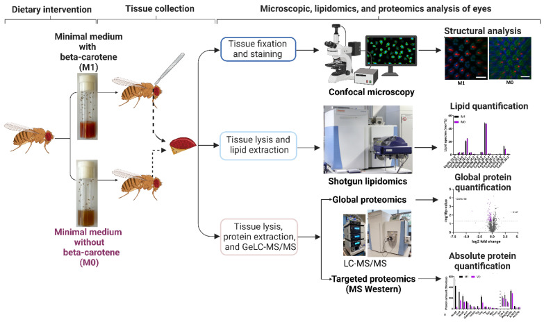
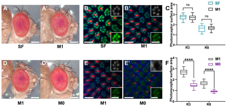
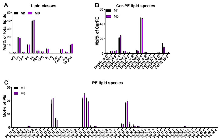
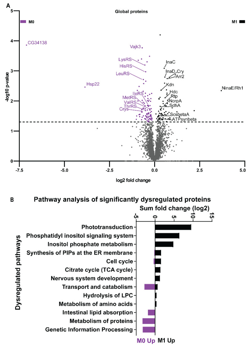
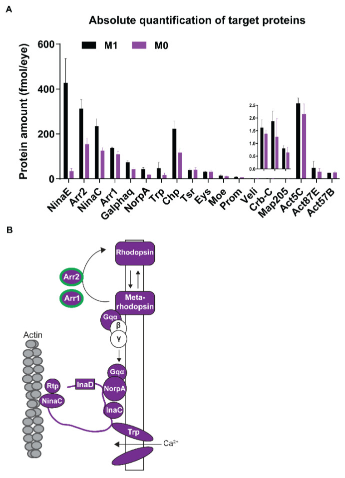
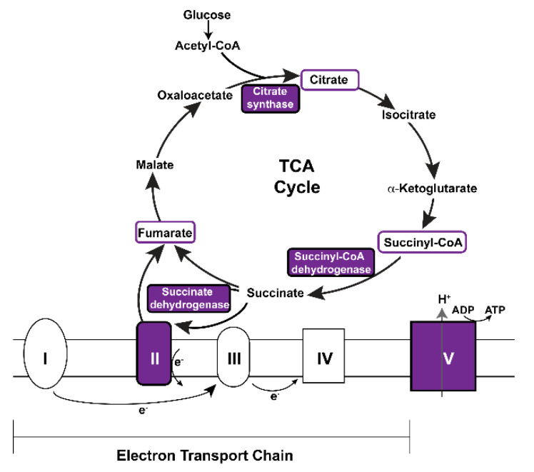
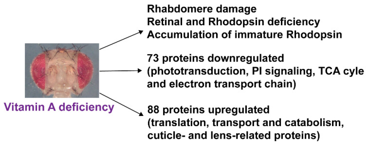
The requirement of vitamin A for the synthesis of the visual chromophore and the light-sensing pigments has been studied in vertebrate and invertebrate model organisms. To identify the molecular mechanisms that orchestrate the ocular response to vitamin A deprivation, we took advantage of the fact that Drosophila melanogaster predominantly requires vitamin A for vision, but not for development or survival. We analyzed the impacts of vitamin A deficiency on the morphology, the lipidome, and the proteome of the Drosophila eye. We found that chronic vitamin A deprivation damaged the light-sensing compartments and caused a dramatic loss of visual pigments, but also decreased the molar abundance of most phototransduction proteins that amplify and transduce the visual signal. Unexpectedly, vitamin A deficiency also decreased the abundances of specific subunits of mitochondrial TCA cycle and respiratory chain components but increased the levels of cuticle- and lens-related proteins. In contrast, we found no apparent effects of vitamin A deficiency on the ocular lipidome. In summary, chronic vitamin A deficiency decreases the levels of most components of the visual signaling pathway, but also affects molecular pathways that are not vision-specific and whose mechanistic connection to vitamin A remains to be elucidated.

Keywords: Drosophila; lipidome; mitochondrion; phototransduction; proteome; retina; retinal; vitamin A.


Conflict of interest statement

The authors declare no conflict of interest. The funders had no role in the design of the study; in the collection, analyses, or interpretation of data; in the writing of the manuscript, or in the decision to publish the results.

Figures

Figure 1
Figure 1
Experimental workflow to analyze the impacts of vitamin A deficiency on the morphology, the lipidome, and the proteome of the Drosophila eye. Schematics were created with BioRender. For details, see text.
Figure 2
Figure 2
Eye and rhabdomere morphology of wild type Drosophila melanogaster raised on nutrient-rich ‘standard’ lab diet (SF), synthetic M1 diet containing a vitamin A precursor, or synthetic M0 diet without vitamin A precursors. (A,A’) Compound eyes of wild type flies that were raised on a ‘standard’ lab diet (SF) or a minimal synthetic diet (M1). Scale bars, 250 µm. (B,B’) Adult retina confocal cross-sections of wild type flies raised on SF or M1 diet. Seven F actin-rich (Phalloidin, green) rhabdomeres are visible in each unit eye (green channel); Rh1 (blue and gray channel) is expressed in the rhabdomeres of ‘outer’ photoreceptors and Rh6 (red) is expressed in the rhabdomeres of ‘inner’ photoreceptors. Scale bars, 10 µm (insets, 5 µm). (C) Quantification of the cross-sectional areas of the rhabdomeres (n = 25 for each condition) for two photoreceptor types (R3: ‘outer’ photoreceptor; R8: ‘inner’ photoreceptor) of flies raised on SF or M1 diet. ns = not significant. (D,D’) Compound eyes of wild type flies that were raised on M1 control diet or M0 diet that lacks vitamin A precursors. Scale bars, 250 µm. (E,E’) Adult retina confocal cross-sections of wild type flies raised on M1 or M0 diet. Seven circular rhabdomeres (Phalloidin, green) are present in each unit eye (green channel). Rh1 (blue and gray channel) is specifically expressed in the rhabdomeres of ‘outer’ photoreceptors and Rh6 (red) is expressed in the rhabdomeres of ‘inner’ photoreceptors in the case of M1 diet. Note the reduced rhabdomere cross-sectional area (green) as well as the abnormal accumulation of Rh1 (blue and gray) and Rh6 (red) outside of the rhabdomeres in the case of M0 diet. Scale bars, 10 µm (insets, 5 µm). (F) Quantification of the cross-sectional areas of the rhabdomeres (n = 25 for each condition) of two different photoreceptor types (R3: ‘outer’ photoreceptor; R8: ‘inner’ photoreceptor) for M1 and M0 diet.
Figure 3
Figure 3
The eye lipidome of wild type Drosophila melanogaster raised on synthetic M1 diet (contains vitamin A precursor) or synthetic M0 diet (lacks vitamin A precursors). (A) Quantification (in mol%) of different lipid classes in the wild type Drosophila eye for M1 diet (control, black) and M0 diet (vitamin A deficiency, purple). (B) Analysis of ocular phospatidylethanolamine ceramide (Cer-PE) lipid species for M1 diet (black) and M0 diet (purple). (C) Analysis of ocular phospatidylethanolamine (PE) lipid species for M1 diet (black) and M0 diet (purple).
Figure 4
Figure 4
The eye proteome of wild type Drosophila melanogaster raised on synthetic M1 diet (contains vitamin A precursor) or synthetic M0 diet (lacks vitamin A precursors). (A) Label-free global eye proteome analysis. The volcano plot shows differentially expressed proteins between M1 diet (black) and M0 diet (purple) in the Drosophila eye. The statistical cut-off (p-value -log10 1.3, equivalent to p < 0.05) is indicated by the dashed line. (B) Pathway enrichment analysis of differentially expressed proteins in the eyes of flies raised on M1 diet or M0 diet. Note that the pathways ‘phototransduction’ and ‘citrate cycle’ were significantly downregulated in the absence of vitamin A, while ‘metabolism of proteins’ was upregulated.
Figure 5
Figure 5
Quantification of major photoreceptor proteins of wild type Drosophila melanogaster raised on synthetic M1 diet or synthetic M0 diet (lacks vitamin A precursors). (A) Targeted absolute quantification of the molar amounts of proteins that play a major role in phototransduction or photoreceptor morphology for M1 diet (black) and M0 diet (purple), N = 3 for each condition. Note the reduced levels of most phototransduction proteins and the structural protein Chp in vitamin A-deficient eyes (purple). Error bar indicates mean ± SD. (B) Vitamin A deficiency significantly decreases the abundances of phototransduction proteins. Note that almost all components are significantly decreased (purple color); Arr1 and Arr2 are regulated on the transcriptional level (green outline).
Figure 6
Figure 6
Vitamin A deficiency decreases the abundances of key components of the mitochondrial TCA cycle and electron transport chain. Purple color indicates significantly decreased proteins or subunits in vitamin A-deficient eyes. For details, see text.
Figure 7
Figure 7
Summary of the morphological and molecular impacts of vitamin A deficiency on the Drosophila melanogaster eye. For details, see text.

Similar articles

Cited by

References

    1. Sommer A. Vitamin a deficiency and clinical disease: An historical overview. J. Nutr. 2008;138:1835–1839. doi: 10.1093/jn/138.10.1835. - DOI - PubMed
    1. Dewett D., Labaf M., Lam-Kamath K., Zarringhalam K., Rister J. Vitamin A deficiency affects gene expression in the Drosophila melanogaster head. G3. 2021;11:jkab297. doi: 10.1093/g3journal/jkab297. - DOI - PMC - PubMed
    1. Saari J.C. Vitamin A and Vision. Subcell. Biochem. 2016;81:231–259. doi: 10.1007/978-94-024-0945-1_9. - DOI - PubMed
    1. von Lintig J. Metabolism of carotenoids and retinoids related to vision. J. Biol. Chem. 2012;287:1627–1634. doi: 10.1074/jbc.R111.303990. - DOI - PMC - PubMed
    1. Nichols R., Pak W.L. Characterization of Drosophila melanogaster rhodopsin. J. Biol. Chem. 1985;260:12670–12674. doi: 10.1016/S0021-9258(17)38924-X. - DOI - PubMed

Publication types

LinkOut - more resources