. 2022 Aug 8;12(8):1091.
doi: 10.3390/biom12081091.

Effect of Human Platelet Lysate as Cultivation Nutrient Supplement on Human Natal Dental Pulp Stem Cell In Vitro Expansion

Affiliations

Effect of Human Platelet Lysate as Cultivation Nutrient Supplement on Human Natal Dental Pulp Stem Cell In Vitro Expansion

Nela Pilbauerova et al. Biomolecules. .

Abstract

Despite several scientific or ethical issues, fetal bovine serum (FBS) remains the standard nutrient supplement in the mesenchymal stem cell cultivation medium. Cell amplification plays an important role in human stem cell therapies. Increasing interest in this field has supported attempts to find suitable human alternatives to FBS for in vitro cell propagation. Human platelet lysate (hPL) has recently been determined as one of them. Our study aimed to evaluate the influence of 2% hPL in the growth medium for in vitro expansion of human natal dental pulp stem cells (hNDP-SCs). The effect was determined on proliferation rate, viability, phenotype profile, expression of several markers, relative telomere length change, and differentiation potential of four lineages of hNDP-SCs. As a control, hNDP-SCs were simultaneously cultivated in 2% FBS. hNDP-SCs cultivated in hPL showed a statistically significantly higher proliferation rate in initial passages. We did not observe a statistically significant effect on mesenchymal stem cell marker (CD29, CD44, CD73, CD90) or stromal-associated marker (CD13, CD166) expression. The cell viability, relative telomere length, or multipotency remained unaffected in hNDP-SCs cultivated in hPL-medium. In conclusion, hPL produced under controlled and standardized conditions is an efficient serum supplement for in vitro expansion of hNDP-SCs.

Keywords: culture medium nutrient supplement; fetal bovine serum; human natal stem cells; human platelet lysate; mesenchymal stem cells; regenerative medicine; stem cell cultivation.


Conflict of interest statement

The authors declare no conflict of interest.

Figures

Figure 1
Figure 1
The rounded spindle-like shape of hNDP-SCs, cultivated for 14 days upon their isolation. Scale bar 50 µm: (a) hNDP-SCs cultivated in the FBS-culture medium; (b) hNDP-SCs cultivated in the hPL-culture medium.
Figure 2
Figure 2
The proliferation rate of hNDP-SCs. The first passage was considered the initial for comparing hNDP-SCs grown in two different media. The data are presented as the mean ± SD. The Shapiro–Wilk test or Kolmogorov–Smirnov test were used for normal distribution evaluations. The statistical significances (** p < 0.01) were calculated using the paired t-test: (a) Cumulative populations doubling; (b) Population doubling time in hours.
Figure 3
Figure 3
Viability of hNDP-SCs cultivated 2% of hPL or FBS supplement measured in the 3rd and 11th passages. The data are presented as the mean ± SD. The Shapiro–Wilk test or Kolmogorov–Smirnov test were used for normal distribution evaluations. The statistical analysis was calculated using the paired t-test. The difference was not statistically significant.
Figure 4
Figure 4
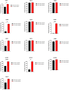
Phenotype profile measured in the 5th passage using flow cytometry. hNDP-SCs from both groups were stained with primary immunofluorescence antibodies conjugated with phycoerythrin (PE) or fluorescein (FITC) against the analyzed CD markers. The percentage of positive cells was determined as a percentage of cells with higher fluorescence intensity than the upper 0.5% isotype immunoglobulin control. Graphs depict the average expression of all analyzed CD markers. The data are presented as the mean ± SD. The Shapiro–Wilk test or Kolmogorov–Smirnov test were used for normal distribution evaluations. The statistical analyses were calculated using the paired t-test (* p < 0.05, *** p < 0.001, **** p < 0.0001).
Figure 5
Figure 5
Immunocytochemical detection of Beta-3-tubulin, the early neuronal marker, in undifferentiated hNDP-SCs harvested in the 7th passage. Most cells were positive for Beta-3-tubulin (red fluorescence). Cell nuclei fluorescent blue. Scale bare 50 µm: (a) hNDP-SCs cultivated in the FBS-culture medium; (b) hNDP-SCs cultivated in the hPL-culture medium.
Figure 6
Figure 6
Immunocytochemical detection of Nestin, the neural progenitor marker, in undifferentiated hNDP-SCs harvested in the 7th passage. Most cells were positive for Nestin (red fluorescence). Cell nuclei fluorescent blue. Scale bare 50 µm: (a) hNDP-SCs cultivated in the FBS-culture medium; (b) hNDP-SCs cultivated in the hPL-culture medium.
Figure 7
Figure 7
Immunocytochemical detection of neurofilaments, the late neuronal marker, in undifferentiated hNDP-SCs harvested in the 7th passage. Most cells were positive for neurofilaments (red fluorescence). Cell nuclei fluorescent blue. Scale bare 50 µm: (a) hNDP-SCs cultivated in the FBS-culture medium; (b) hNDP-SCs cultivated in the hPL-culture medium.
Figure 8
Figure 8
Immunocytochemical detection of Nanog, marker playing a role in ESC pluripotency, maintenance, and self-renewal, in undifferentiated hNDP-SCs harvested in the 7th passage. Most cells were positive for Nanog (red fluorescence). Cell nuclei fluorescent blue. Scale bare 50 µm: (a) hNDP-SCs cultivated in the FBS-culture medium; (b) hNDP-SCs cultivated in the hPL-culture medium.
Figure 9
Figure 9
Average relative telomere length measured between 3rd and 14th passages using qPCR. Both groups of hNDP-SCs experienced the shortening of relative telomere length in the 14th passage (* p < 0.05), but it was more noticeable in the case of hNDP-SCs cultivated in the medium with 2% FBS. The data are presented as the mean ± SD. The Shapiro–Wilk test or Kolmogorov–Smirnov test were used for normal distribution evaluations. The statistical analyses were calculated using the paired t-test.
Figure 10
Figure 10
Detection of chondrogenic differentiation in the extracellular matrix of hNDP-SCs cultivated in the FBS-culture medium. Scale bar 50 µm: (ac) After 3-week cultivation in chondrogenic differentiation medium; (df) undifferentiated hNDP-SCs; (a,d) immunocytochemical detection of collagen type II (red fluorescence); (b,e) histological detection of collagen and procollagen after blue Masson’s trichrome stain (blue areas); (c,f) histological detection of acid mucopolysaccharides after Alcian blue stain (turquoise areas).
Figure 10
Figure 10
Detection of chondrogenic differentiation in the extracellular matrix of hNDP-SCs cultivated in the FBS-culture medium. Scale bar 50 µm: (ac) After 3-week cultivation in chondrogenic differentiation medium; (df) undifferentiated hNDP-SCs; (a,d) immunocytochemical detection of collagen type II (red fluorescence); (b,e) histological detection of collagen and procollagen after blue Masson’s trichrome stain (blue areas); (c,f) histological detection of acid mucopolysaccharides after Alcian blue stain (turquoise areas).
Figure 11
Figure 11
Detection of chondrogenic differentiation in the extracellular matrix of hNDP-SCs cultivated in the hPL-culture medium. Scale bar 50 µm: (ac) After 3-week cultivation in chondrogenic differentiation medium; (df) undifferentiated hNDP-SCs; (a,d) immunocytochemical detection of collagen type II (red fluorescence); (b,e) histological detection of collagen and procollagen after blue Masson’s trichrome stain (blue areas); (c,f) histological detection of acid mucopolysaccharides after Alcian blue stain (turquoise areas).
Figure 12
Figure 12
Detection of osteogenic differentiation in the extracellular matrix of hNDP-SCs cultivated in the FBS-culture medium. Scale bar 50 µm: (ac) After 3-week cultivation in osteogenic differentiation medium; (df) undifferentiated hNDP-SCs; (a,d) immunocytochemical detection of osteocalcin (brown or rusty areas); (b,e) histological detection of calcium phosphate deposits after von Kossa stain (dark brown or black spots); (c,f) histological detection of calcium deposits after Alizarin Red stain (red areas).
Figure 13
Figure 13
Detection of osteogenic differentiation in the extracellular matrix of hNDP-SCs cultivated in the hPL-culture medium. Scale bar 50 µm: (ac) After 3-week cultivation in osteogenic differentiation medium; (df) undifferentiated hNDP-SCs; (a,d) immunocytochemical detection of osteocalcin (brown or rusty areas); (b,e) histological detection of calcium phosphate deposits after von Kossa stain (dark brown or black spots); (c,f) histological detection of calcium deposits after Alizarin Red stain (red areas).
Figure 14
Figure 14
Detection of adipose droplets and vacuoles in the extracellular matrix of hNDP-SCs after 4-week cultivation in adipogenic differentiation medium. After red oil staining, the adipose vacuoles are revealed as red areas. Scale bar 50 µm: (a,b) hNDP-SCs cultivated in the FBS-culture medium; (c,d) hNDP-SCs cultivated in the hPL-culture medium; (a,c) phase contrast optical microscope phase contrast microscope; (b,d) inverted optical microscope.

Similar articles

Cited by

References

    1. Mhaske S., Yuwanati M.B., Mhaske A., Ragavendra R., Kamath K., Saawarn S. Natal and neonatal teeth: An overview of the literature. ISRN Pediatr. 2013;2013:956269. doi: 10.1155/2013/956269. - DOI - PMC - PubMed
    1. Leung A.K., Robson W.L. Natal teeth: A review. J. Natl. Med. Assoc. 2006;98:226–228. - PMC - PubMed
    1. Leung A.K. Natal teeth. Am. J. Dis. Child. 1986;140:249–251. doi: 10.1001/archpedi.1986.02140170075033. - DOI - PubMed
    1. Seminario A.L., Ivancaková R. Natal and neonatal teeth. Acta Med. 2004;47:229–233. doi: 10.14712/18059694.2018.96. - DOI - PubMed
    1. Kates G.A., Needleman H.L., Holmes L.B. Natal and neonatal teeth: A clinical study. J. Am. Dent. Assoc. 1984;109:441–443. doi: 10.14219/jada.archive.1984.0415. - DOI - PubMed

Publication types