. 2022 Aug 12;12(8):1108.
doi: 10.3390/biom12081108.

Translocation of Distinct Alpha Synuclein Species from the Nucleus to Neuronal Processes during Neuronal Differentiation

Affiliations

Translocation of Distinct Alpha Synuclein Species from the Nucleus to Neuronal Processes during Neuronal Differentiation

Katharina Pieger et al. Biomolecules. .

Abstract

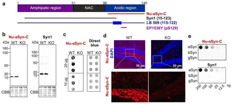
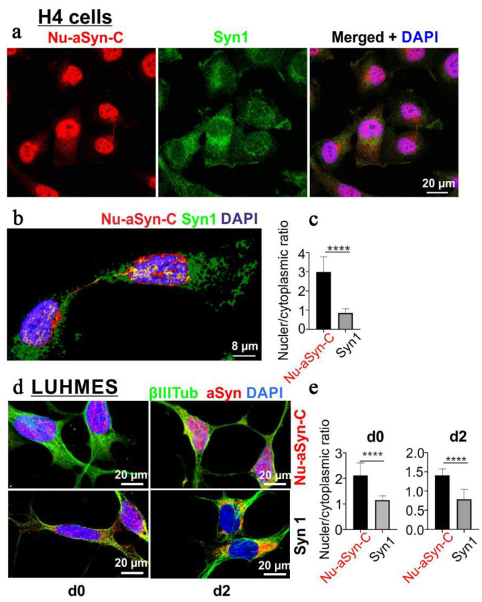
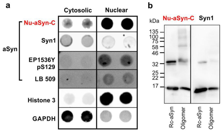
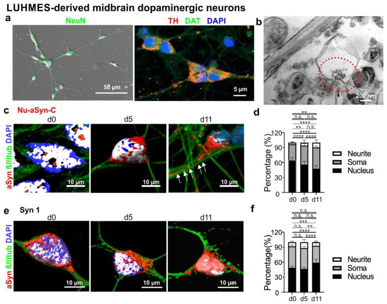
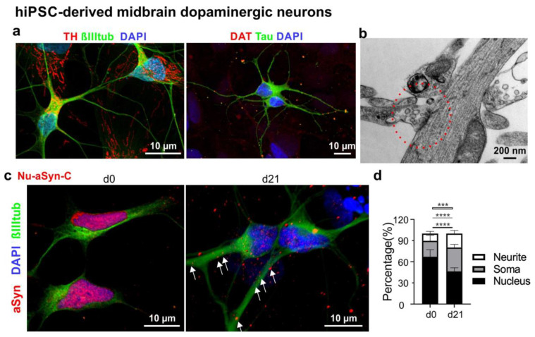
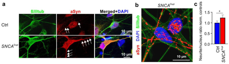
Alpha synuclein (aSyn) and its aggregation are crucial for the neurodegeneration of Parkinson's disease (PD). aSyn was initially described in the nucleus and presynaptic nerve terminals. However, the biology of nuclear aSyn and the link of aSyn between subcellular compartments are less understood. Current knowledge suggests the existence of various aSyn species with distinct structural and biochemical properties. Here, we identified a C-terminal-targeting aSyn antibody (Nu-aSyn-C), which has a high immunoaffinity towards aSyn in the nucleus. Comparing the Nu-aSyn-C antibody to aSyn antibodies developed against phosphorylated or aggregated forms, we observed that nuclear aSyn differs from cytosolic aSyn by an increased phosphorylation and assembly level in proliferating cells. Employing Nu-aSyn-C, we characterized aSyn distribution during neuronal differentiation in midbrain dopaminergic neurons (mDANs) derived from human-induced pluripotent stem cells (hiPSCs) and Lund human mesencephalic cells, and in primary rat hippocampal neurons. We detected a specific translocation pattern of aSyn during neuronal differentiation from the nucleus to the soma and finally to neuronal processes. Interestingly, a remarkable shift of Nu-aSyn-C-positive species towards neurites was detected in hiPSC mDANs from a PD patient carrying aSyn gene duplication. Together, our results reveal distinct nuclear and cytosolic aSyn species that redistribute during neuronal differentiation-a process that is altered in PD-derived neurons.

Keywords: Parkinson’s disease; SNCA duplication; alpha synuclein; nuclear alpha synuclein.


Conflict of interest statement

The authors declare no conflict of interest.

Figures

Figure 1
Figure 1
Nu-aSyn-C antibody is specific to aSyn. (a) Structure of human aSyn and antibodies against different aSyn epitopes applied in this study. (b) Denaturing WB analysis of brain lysates of WT and aSyn KO mice using Nu-aSyn-C and Syn1 antibodies demonstrates specificity of both antibodies to aSyn in WT mice. (c) Non-denaturing dot blot analysis of WT and aSyn KO mouse brains reveals immunosignals in WT mice only. An amount of 10–20 µg of total protein extract was loaded for WB (b) or dot blot (c). Loading of the total protein extract was controlled by CBB (WB, (b)) or using direct blue 71 dye (dot blot, (c)). (d) IHC analysis of WT and KO brain sections using the Nu-aSyn-C antibody shows specific immunosignals in WT brains only (example images from the hippocampus in d; example images from the cortex in Figure S1. (e) Dot blot analysis of human recombinant a-, b-, and gSyn using Nu-aSyn-C and Syn1 antibodies. Both antibodies do not cross-react with b- and gSyn.
Figure 2
Figure 2
Labelling of aSyn with Nu-aSyn-C and Syn1 antibodies in proliferating H4 and LUHMES cells (d0), as well as in LUHMES cells after 2 days of differentiation. Nu-aSyn-C immunosignals are predominantly present in the nucleus, whereas Syn1 immunosignals are detected to a larger extent in the cytosol. (a) ICC analysis of aSyn distribution in H4 cells detected by Nu-aSyn-C and Syn1 antibodies. (b) A 3D reconstruction of representative proliferating H4 cells. (c) Ratio of nuclear versus cytoplasmic immunosignal intensity detected by Nu-aSyn-C and Syn1 antibodies, respectively. Data represent the mean ± SD from 35 cells for each antibody. Statistics: Mann–Whitney test; **** p < 0.001. (d) aSyn distribution patterns recognized by Nu-aSyn-C and Syn1 antibodies, respectively, in proliferating (d0) and differentiating LUHMES cells (d2). (e) Quantification of the ratios of nuclear and cytoplasmic immunosignal intensity. Data represent the mean ± SD from 11–21 cells for each antibody. Statistics: Mann–Whitney test; **** p < 0.001.
Figure 3
Figure 3
(a) Dot blot analysis of enriched cytosolic and nuclear fractions from H4 cells using different anti-aSyn antibodies, including Nu-aSyn-C, Syn1, EP1536Y (specific for phosphorylated aSyn on serine 129), and LB 509 (recognizing aggregated aSyn). GAPDH and histone 3 serve as protein markers of the cytosol and the nucleus, respectively. In H4 cells, nuclear aSyn exhibits a higher phosphorylated and aggregated status than cytosolic aSyn. (b) WB analysis of human recombinant aSyn (Rc-aSyn) and preformed aSyn oligomers. The Nu-aSyn-C antibody preferentially recognizes higher-molecular-weight oligomers.
Figure 4
Figure 4
Characterization of mDANs differentiated from LUHMES cells. (a) Representative neurons differentiated for 5 days show the expression of the neuronal marker NeuN (left, green) and dopaminergic neuronal markers TH (red) and DAT (green). (b) Representative EM image with a synaptic site (red circle) in LUHMES cells differentiated for 35 days. (c,e) Representative 3D reconstructed fluorescence images of LUHMES cells at the proliferating stage (d0), and differentiated for 5 (d5) and 11 days (d11) labelled with either the Nu-aSyn-C (c) or Syn1 antibody (e), βIIItub and DAPI. (d,f) Quantification of aSyn distribution (in %) in the nucleus, soma, and neurites, labeled with either the Nu-aSyn-C (d) or Syn1 (f) antibody. In the 3D reconstructed images (c,e), nuclear localization of aSyn is depicted in white due to the special morphology of LUHMES cell bodies characterized by a close arrangement of the nucleus and the surrounding soma. The Nu-aSyn-C antibody detects a strong localization of aSyn in the nucleus of proliferating cells (d0). Upon differentiation (d5–d11), aSyn signal intensity in the soma and neurites significantly increases. This shift of the aSyn signal revealed by the Nu-aSyn-C antibody is much less visible using the Syn1 antibody. For quantification (d,f), data represent the mean ± SD from 9–19 cells of each time point measured in three independent experiments. Statistics: Two-way ANOVA, Tukey’s multiple comparisons test, n.s.: not significant, ** p < 0.005, *** p < 0.0005, and **** p < 0.0001. Black lines: comparisons of nuclear signal intensity; grey lines: comparisons of soma signal intensity; white lines: comparisons of neurite signal intensity.
Figure 5
Figure 5
Characterization of mDANs differentiated from hiPSCs. (a) Representative neurons differentiated for 21 days show the presence of the neuronal markers βIIItub and Tau (green) as well as the dopaminergic neuronal markers TH and DAT (red). (b) Representative EM image of a putative synaptic site (red circle) with synaptic vesicles in hiPSC-derived neurons differentiated for 21 days. (c) Representative immunofluorescence images of proliferating smNPCs (d0) and neurons differentiated for 21 days (d21) labelled with the Nu-aSyn-C antibody, an anti-βIIItub antibody, and DAPI. Immunosignals for aSyn and DAPI were reconstructed in 3D to demonstrate the distribution pattern of aSyn. (d) Quantification of aSyn distribution (in %) in the nucleus, soma and neurites. The Nu-aSyn-C antibody detects aSyn in the nucleus of proliferating cells (d0). Upon differentiation (d21), aSyn signal intensity significantly decreases in the nucleus and increases in the soma and neurites. For quantification, data represent the mean ± SD of >16 cells of each time point measured in three independent experiments. Two hiPSC lines (UKERi33Q-R1-006 and UKERi1E4-R1-003) from two healthy individuals were used. Statistics: Two-way ANOVA, Tukey’s multiple comparisons test, *** p < 0.0005, and **** p < 0.0001. Black lines: comparisons of nuclear signal intensity; grey lines: comparisons of soma signal intensity; white lines: comparisons of neuritic intensity.
Figure 6
Figure 6
ICC analysis of aSyn distribution in mDANs from healthy controls (Ctrl) and a SNCADupl patient. (a) Representative immunofluorescence images of mDANs differentiated for 21 days labelled with the Nu-aSyn-C antibody, anti-βIIItub, and DAPI. The corresponding TH staining is shown in Figure S5. (b) Representative reconstructed 3D image highlights intense aSyn immunosignals along the neurites of SNCADupl mDANs. (c) Quantification of neurite/nucleus ratios of aSyn immunosignal intensity in SNCADupl mDANs normalized to the average ratio in control-derived neurons. For quantification, data represent the mean ± SD of >10 neurons of each hiPSC line in three independent experiments. Three hiPSC cell lines from three healthy individuals (UKERi33Q-R1-006, UKERiO3H-R1-001, and UKERi82A-S1-017) and two hiPSC lines (CSC-1A and CSC-1D) from the SNCADupl patient were used. Statistics: Two-way ANOVA, Tukey’s multiple comparisons test * p < 0.05.

Similar articles

Cited by

References

    1. Iwai A., Masliah E., Yoshimoto M., Ge N., Flanagan L., de Silva H.A., Kittel A., Saitoh T. The precursor protein of non-A beta component of Alzheimer’s disease amyloid is a presynaptic protein of the central nervous system. Neuron. 1995;14:467–475. doi: 10.1016/0896-6273(95)90302-X. - DOI - PubMed
    1. Iwai A. Properties of NACP/alpha-synuclein and its role in Alzheimer’s disease. Biochim. Biophy. Acta. 2000;1502:95–109. doi: 10.1016/S0925-4439(00)00036-3. - DOI - PubMed
    1. Klein C., Westenberger A. Genetics of Parkinson’s disease. Cold Spring Harb. Perspect. Med. 2012;2:a008888. doi: 10.1101/cshperspect.a008888. - DOI - PMC - PubMed
    1. Baba M., Nakajo S., Tu P.H., Tomita T., Nakaya K., Lee V.M., Trojanowski J.Q., Iwatsubo T. Aggregation of alpha-synuclein in Lewy bodies of sporadic Parkinson’s disease and dementia with Lewy bodies. Am. J. Pathol. 1999;152:879–884. - PMC - PubMed
    1. Spillantini M.G., Schmidt M.L., Lee V.M., Trojanowski J.Q., Jakes R., Goedert M. Alpha-synuclein in Lewy bodies. Nature. 1997;388:839–840. doi: 10.1038/42166. - DOI - PubMed

Publication types

Substances