. 2022 Aug 12;12(8):1111.
doi: 10.3390/biom12081111.

Structure-Activity Relationship of the Dimeric and Oligomeric Forms of a Cytotoxic Biotherapeutic Based on Diphtheria Toxin

Affiliations

Structure-Activity Relationship of the Dimeric and Oligomeric Forms of a Cytotoxic Biotherapeutic Based on Diphtheria Toxin

Marcin Mielecki et al. Biomolecules. .

Abstract

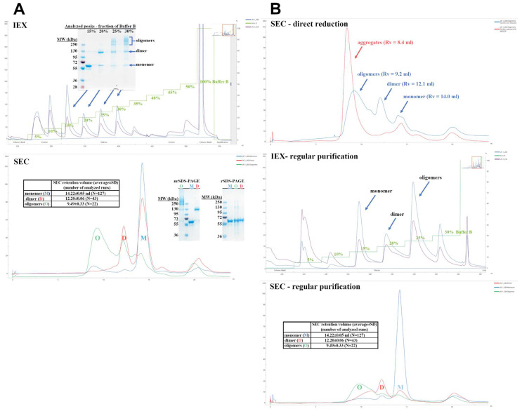
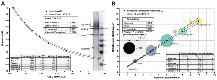
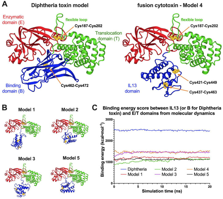
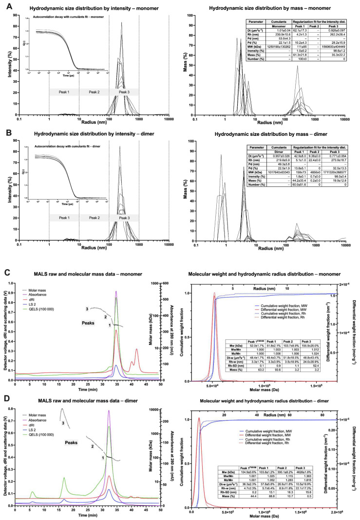
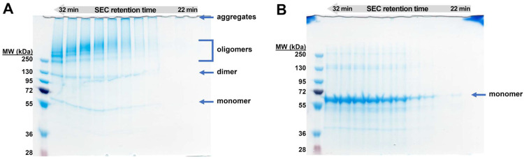
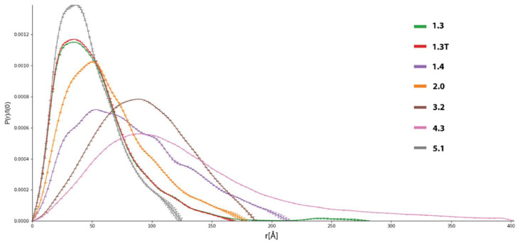
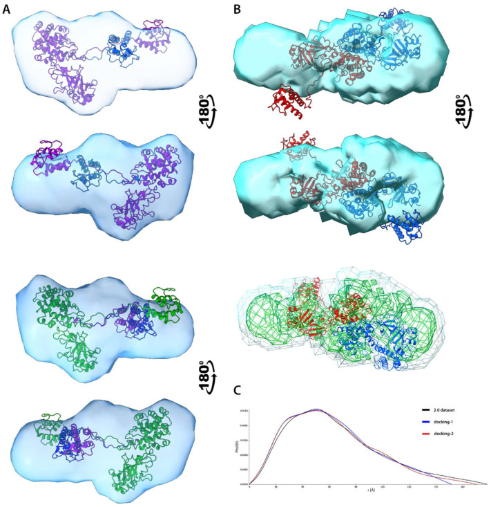
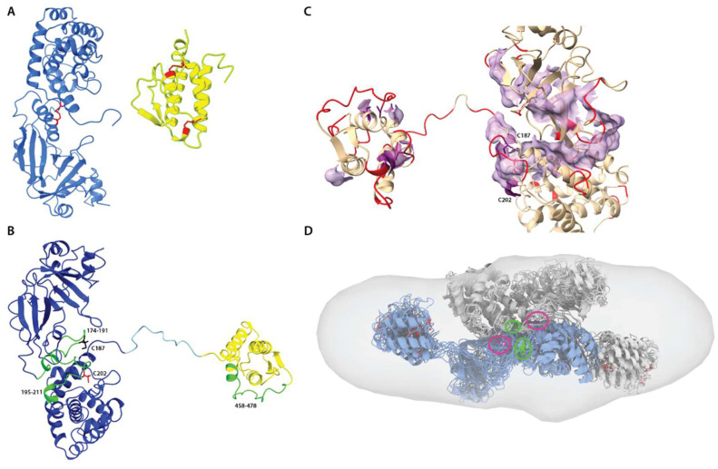
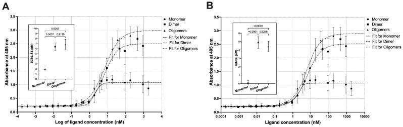
Protein aggregation is a well-recognized problem in industrial preparation, including biotherapeutics. These low-energy states constantly compete with a native-like conformation, which is more pronounced in the case of macromolecules of low stability in the solution. A better understanding of the structure and function of such aggregates is generally required for the more rational development of therapeutic proteins, including single-chain fusion cytotoxins to target specific receptors on cancer cells. Here, we identified and purified such particles as side products of the renaturation process of the single-chain fusion cytotoxin, composed of two diphtheria toxin (DT) domains and interleukin 13 (IL-13), and applied various experimental techniques to comprehensively understand their molecular architecture and function. Importantly, we distinguished soluble purified dimeric and fractionated oligomeric particles from aggregates. The oligomers are polydisperse and multimodal, with a distribution favoring lower and even stoichiometries, suggesting they are composed of dimeric building units. Importantly, all these oligomeric particles and the monomer are cystine-dependent as their innate disulfide bonds have structural and functional roles. Their reduction triggers aggregation. Presumably the dimer and lower oligomers represent the metastable state, retaining the native disulfide bond. Although significantly reduced in contrast to the monomer, they preserve some fraction of bioactivity, manifested by their IL-13RA2 receptor affinity and selective cytotoxic potency towards the U-251 glioblastoma cell line. These molecular assemblies probably preserve structural integrity and native-like fold, at least to some extent. As our study demonstrated, the dimeric and oligomeric cytotoxin may be an exciting model protein, introducing a new understanding of its monomeric counterpart's molecular characteristics.

Keywords: IL-13; LC/MS; MALS; SAXS; biotherapeutics; cytotoxin; diphtheria toxin; disulfide bond; inclusion bodies; protein oligomerization; refolding.


Conflict of interest statement

The authors declare no conflict of interest. The funders had no role in the design of the study, in the collection, analyses, or interpretation of data, in the writing of the manuscript, or in the decision to publish the results.

Figures

Figure 1
Figure 1
IEX and SEC chromatography of the cytotoxin. (A). IEX chromatogram with the recorded absorbance (254 nm, purple; 280 nm, blue) and conductivity (green) traces presents the initial separation of the protein dialysate into monomer, dimer, and oligomers. The peak fractions were resolved by non-reducing SDS-PAGE, revealing electrophoretic mobility of the monomer and dimer protein bands and a ladder of bands for the oligomer fractions. SEC chromatograms of the separated IEX peak fractions with the overlaid 280 nm traces are presented for the monomer (blue), dimer (red), and oligomers (green). The table inlet contains average elution volumes for each molecular state. The apex fractions were resolved by non-reducing and reducing SDS-PAGE. (B). The cytotoxin recovery from the oligomeric fractions by two tested approaches, direct reduction and regular purification protocol with IEX and SEC chromatography. Analytical SEC of the reduction substrates (blue) and products (red)—shifting the elution volume (VSEC) for the monomer, dimer, and oligomers toward column void volume (about 8.0 mL) indicates aggregation. IEX chromatogram of the solubilized and refolded oligomers, separated into monomers, dimers, and oligomers. SEC chromatograms of the separated IEX peak fractions with the overlaid 280 nm traces are presented for the monomer (blue), dimer (red), and oligomers (green). The table inlet contains average elution volumes for each molecular state. X axes indicate elution volume (mL).
Figure 2
Figure 2
Determination of oligomers stoichiometry in non-reducing SDS-PAGE gels by analyzing relative front parameter (Rf). (A). Exemplary non-linear regression of the standard molecular weights plotted against their normalized relative fronts. The power model parameters were used to derive an apparent molecular weight of the cytotoxin bands, and their stoichiometries averaged for several gel paths. (B). The linear correlation between Rf-estimated and assumed stoichiometries for the dimer and oligomers. The Rf value for each band was also compared to the theoretical stoichiometries by the one-sample t-test, and the resulting p-values are displayed on the bubble plot. Each band was compared to three or four most probable values. Dotted lines represent the 95% confidence intervals (CI) for the regression models.
Figure 3
Figure 3
Molecular dynamics simulations for the diphtheria toxin and the fusion cytotoxin monomer. Presented are the ribbon representations of the last snapshots of the 20 ns MD. (A). Diphtheria toxin model compared to the cytotoxin Model 4, the one with the highest average DT390 and IL-13 interdomain binding energy score. (B). Structures of four other constructed models. (C). Trajectories of DT and DT390-IL-13 models as “binding energy” parameter in the function of time.
Figure 4
Figure 4
Results of DLS and MALS measurements for monomeric and dimeric fractions of DT390-IL-13. (A) DLS of monomeric fractions. (B) DLS of dimeric fraction. (C) MALS of monomeric fractions. (D) MALS of dimeric fraction.
Figure 5
Figure 5
SDS-PAGE analysis in non-reducing (A) and reducing (B) conditions of the consecutive SEC-MALS oligomer fractions with the elution time range of 22–32 min. An amount of 10 µg was loaded into each well. The average mass distribution over the analyzed fractions in the non-reducing conditions is inversely proportional to the SEC-MALS elution time, as expected. The higher the molecular weights, the more unstable the cytotoxin. The oligomeric fractions additionally contain monomer, dimer, and aggregates. The efficient oligomers monomerization was observed in the reducing conditions.
Figure 6
Figure 6
P(r) functions for selected datasets extracted from initial SEC-SAXS data. All these datasets were selected for further analysis.
Figure 7
Figure 7
SAXS structural modelling of DT390-IL-13. (A). Alignment of atomic models representing 2-state (top) and 3-state (bottom) models onto ED reconstruction. Each conformer is depicted using a different color. (B). Alignment of atomic model of best-scoring dimer onto ED reconstruction, each monomer is depicted using a different color (top). Layers of different electron density showing coverage of polypeptide chain (bottom). (C). P(r) functions for both original and 2.0 dataset (representing a dimer)and two selected dimers created by the FoXSDock.
Figure 8
Figure 8
Disulfide mapping and protein-protein recognition. (A). Native disulfide bridges (red) in DT390 domains (blue) and IL-13 (yellow). (B). Cysteine-containing peptides identified by MS (green). Reduced C187 and C202 residues are colored black and red, respectively. (C). Fragment of DT390 with flexible residues colored red and surface of the major hydrophobic path. Residues C187 and C202 are highlighted in purple and belong to the hydrophobic path. (D). Fitting conformational assemblies of DT390-IL-13 dimer to ED model. Each “monomer” is a structural alignment of 10 most probable conformers, and both molecules are manually docked to achieve proximity between C187 and C202 residues highlighted in green and purple, respectively.
Figure 9
Figure 9
Non-linear regression of the ELISA experimental data for the titration of IL13RA2 receptor with the cytotoxin monomer, dimer, and oligomers. Two models were used: (A) sigmoidal, to derive EC50, and (B) quadratic, to derive apparent Kd values. The inlet graphs present the fit parameters, EC50 or Kd, and p-values for the pairwise comparisons by one-way ANOVA.
Figure 10
Figure 10
The viability of U-251 glioblastoma cell line in presence of the cytotoxin monomer (M), dimer (D), and oligomers (F4-E6), measured by the MTS assay, in three concentrations, 0.1, 1.0, and 10.0 ng (which correspond to 1.8, 18.1, and 181.3 pM concentration, respectively). Significant differences are indicated as: for * p < 0.05, ** p < 0.01 and **** for p < 0.001.

Similar articles

Cited by

References

    1. Zahaf N.-I., Schmidt G. Bacterial Toxins for Cancer Therapy. Toxins. 2017;9:236. doi: 10.3390/toxins9080236. - DOI - PMC - PubMed
    1. Peigneur S., Tytgat J. Toxins and drug discovery. Toxins. 2018;10:126. doi: 10.3390/toxins10030126. - DOI - PMC - PubMed
    1. Garashchenko B.L., Korsakova V.A., Yakovlev R.Y. Radiopharmaceuticals Based on Alpha Emitters: Preparation, Properties, and Application. Phys. At. Nucl. 2018;81:1515–1525. doi: 10.1134/S1063778818100071. - DOI
    1. Pagliaro L.C., Liu B., Munker R., Andreeff M., Freireich E.J., Scheinberg D.A., Rosenblum M.G. Humanized M195 monoclonal antibody conjugated to recombinant gelonin: An anti-CD33 immunotoxin with antileukemic activity. Clin. Cancer Res. 1998;4:1971–1976. - PubMed
    1. Chandramohan V., Sampson J.H., Pastan I., Bigner D.D. Toxin-Based Targeted Therapy for Malignant Brain Tumors. Clin. Dev. Immunol. 2012;2012:480429. doi: 10.1155/2012/480429. - DOI - PMC - PubMed

Publication types