Review
. 2022 Aug 3;11(8):1516.
doi: 10.3390/antiox11081516.

Redox Homeostasis in Ocular Tissues: Circadian Regulation of Glutathione in the Lens?

Affiliations
Review

Redox Homeostasis in Ocular Tissues: Circadian Regulation of Glutathione in the Lens?

Julie C Lim et al. Antioxidants (Basel). .

Abstract

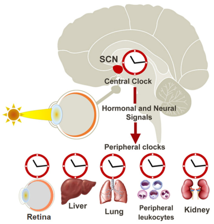
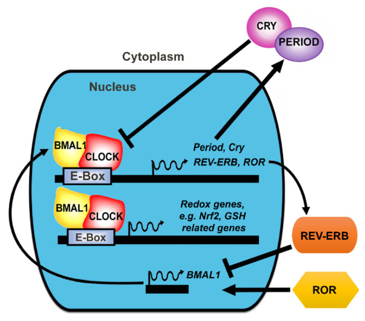
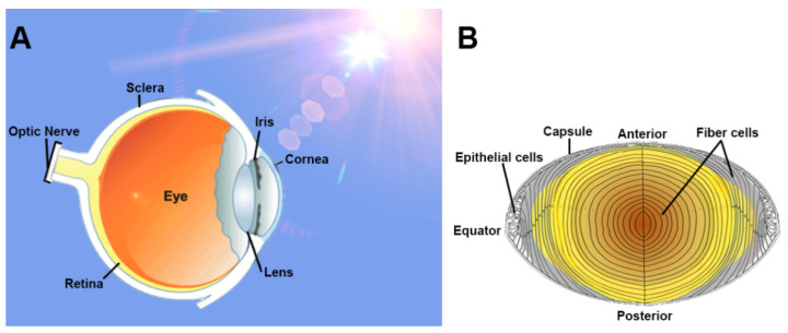
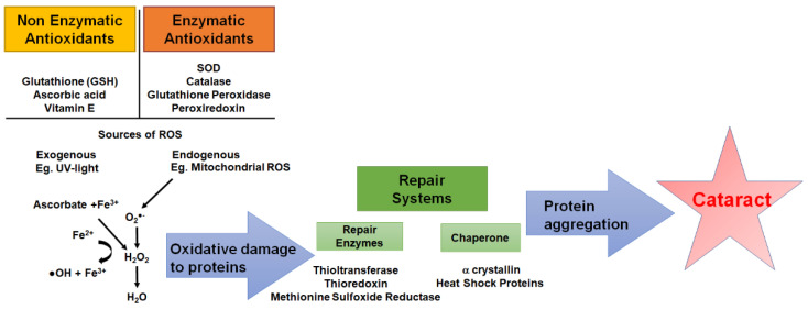
Accumulating evidence in tissues suggests an interconnection between circadian clocks and redox regulation. Diurnal variations in antioxidant levels, circadian rhythms of antioxidant enzyme activity, and differences in oxidative stress markers at different times of the day all indicate that oxidative stress responses follow a circadian rhythm. Disruptions of circadian rhythms are linked to a number of age-related diseases, including those in the eye. Typically, ocular tissues contain a robust antioxidant defence system to maintain redox balance and minimise oxidative stress and damage. The lens, in particular, contains remarkably high levels of the antioxidant glutathione (GSH). However, with advancing age, GSH levels deplete, initiating a chain of biochemical events that ultimately result in protein aggregation, light scattering, and age-related cataracts. While there is evidence that the lens exhibits circadian rhythms in the synthesis and release of melatonin, little is known about the regulation or function of timekeeping mechanisms in the lens. Since circadian rhythms are disrupted with age, and the depletion of GSH in the lens is a known initiating factor in the development of age-related cataracts, understanding the mechanisms involved in regulating GSH levels may lead to the future development of approaches to manipulate the clock to restore GSH levels and redox balance in the lens, and protect the lens from cataracts.

Keywords: cataract; circadian rhythms; glutathione; lens.


Conflict of interest statement

The authors declare no conflict of interest.

Figures

Figure 1
Figure 1
The mammalian circadian clock network. The mammalian circadian clock network consists of the central clock located in the suprachiasmatic nucleus (SCN) of the hypothalamus and peripheral oscillators present in virtually all cell types. Light activates a specific group of photoreceptors in the retina that are connected to the central SCN clock, which synchronises and entrains peripheral circadian clocks via neural and endocrine pathways.
Figure 2
Figure 2
The transcriptional-translational feedback loop. At the molecular level, circadian rhythms are generated by the rhythmic expression of clock genes and the proteins they encode. CLOCK and BMAL1 heterodimers activate transcription of period (Per) and cryptochrome (Cry) genes. PER and CRY proteins, in turn, inhibit their own expression by repressing CLOCK/BMAL1 activity. BMAL1/CLOCK also controls the rhythmic expression of REV-ERBα, which feeds back to suppress BMAL1 transcription. The expression of retinoic acid-related orphan receptor (RORα) is also controlled by BMAL1/CLOCK, which feeds back to activate BMAL1 transcription. BMAL1/CLOCK also activates the transcription of clock-controlled genes such as Nrf2- and GSH-related genes. This feedback loop generates ~24 h oscillations of clock protein levels and activity, which is translated into circadian control of behaviour and physiology.
Figure 3
Figure 3
The lens: function and structure. (A) Light enters the eye through the transparent cornea and lens, which refracts light to focus onto the retina. Light information is converted into electrical impulses, which are sent to the primary visual cortex of the brain via the optic nerve. The lens is positioned in the anterior region of the eye and is therefore vulnerable to damage from reactive oxygen species (ROS) generated by UV radiation. (B) Schematic diagram of the human lens revealing the epithelial monolayer from where epithelial cells proliferate and differentiate at the equatorial zone to form differentiating fibre cells in the outer cortex. Fibre cells progressively lose their organelles, such as mitochondria and nuclei, as they become internalised, creating an inherent age gradient that encapsulates all stages of fibre cell differentiation throughout the lifetime of an individual.
Figure 4
Figure 4
Schematic of the antioxidant defence systems of the lens. To protect against exogenous and endogenous sources of reactive oxygen species (ROS), the lens utilises nonenzymatic antioxidants such as glutathione (GSH), ascorbic acid, and vitamin E along with enzymatic antioxidants such as superoxide dismutase (SOD), which neutralises the superoxide radical (O2-•) and catalase, glutathione peroxidase (GPx) and peroxiredoxins, which neutralises H2O2 into H2O and molecular oxygen (O2). Repair systems such as the repair enzymes thioltransferase, thioredoxin, methionine sulfoxide reductase, and chaperone proteins work to repair oxidised proteins. However, depletion of antioxidant capacity coupled with a failure to repair damaged proteins results in protein aggregation, increased light scattering, and ultimately cataract formation.

Similar articles

Cited by

References

    1. Takahashi J.S. Transcriptional architecture of the mammalian circadian clock. Nat. Rev. Genet. 2016;18:164–179. doi: 10.1038/nrg.2016.150. - DOI - PMC - PubMed
    1. Xie Y., Tang Q., Chen G., Xie M., Yu S., Zhao J., Chen L. New Insights Into the Circadian Rhythm and Its Related Diseases. Front. Physiol. 2019;10:682. doi: 10.3389/fphys.2019.00682. - DOI - PMC - PubMed
    1. Siu A.W., Maldonado M., Hidalgo M.S., Tan D.-X., Reiter R.J. Protective effects of melatonin in experimental free radical-related ocular diseases. J. Pineal Res. 2006;40:101–109. doi: 10.1111/j.1600-079X.2005.00304.x. - DOI - PubMed
    1. Felder-Schmittbuhl M.-P., Buhr E.D., Dkhissi-Benyahya O., Hicks D., Peirson S.N., Ribelayga C.P., Sandu C., Spessert R., Tosini G. Ocular Clocks: Adapting Mechanisms for Eye Functions and Health. Investig. Opthalmol. Vis. Sci. 2018;59:4856–4870. doi: 10.1167/iovs.18-24957. - DOI - PMC - PubMed
    1. Giblin F.J. Glutathione: A Vital Lens Antioxidant. J. Ocul. Pharmacol. Ther. 2000;16:121–135. doi: 10.1089/jop.2000.16.121. - DOI - PubMed

LinkOut - more resources