

Comparative Proteomic Assessment of Normal vs. Polyhydramnios Amniotic Fluid Based on Computational Analysis
- PMID: 36009368
- PMCID: PMC9404943
- DOI: 10.3390/biomedicines10081821
Comparative Proteomic Assessment of Normal vs. Polyhydramnios Amniotic Fluid Based on Computational Analysis
Abstract
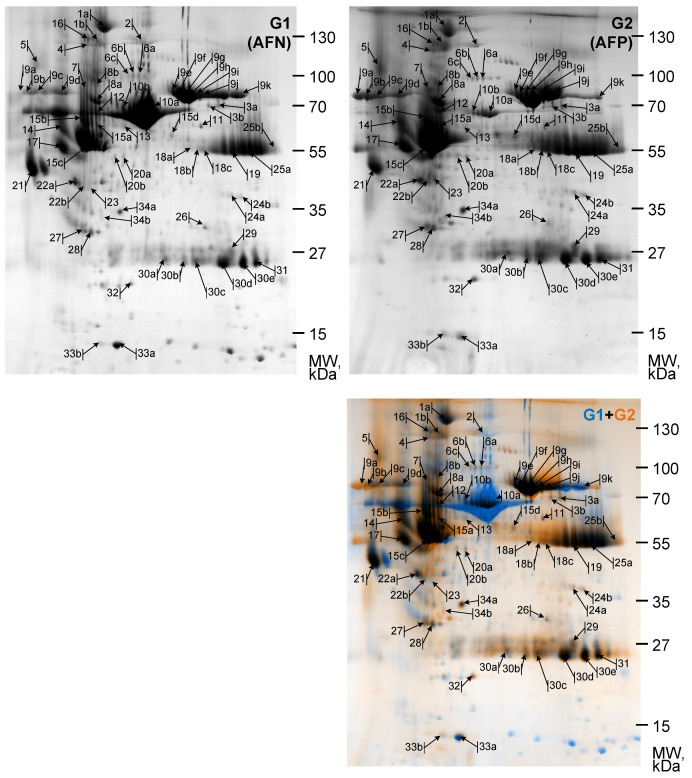
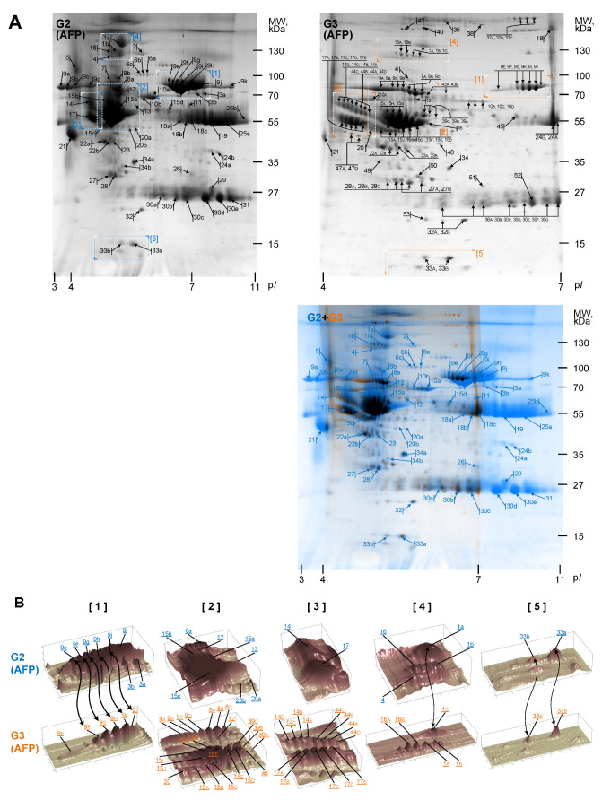
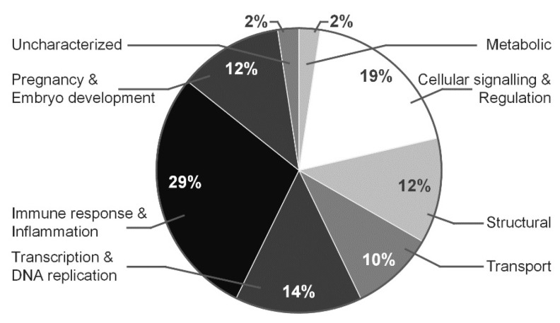
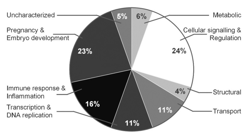
Mass spectrometry-based proteomics have become a valued tool for conducting comprehensive analyses in amniotic fluid samples with pathologies. Our research interest is the finding and characterization of proteins related to normal vs. polyhydramnios (non-immune hydrops) pregnancy. Proteomic analysis was performed on proteins isolated from fresh amniotic fluid samples. Proteins were fractionated by 2DE using a different pI range (pI 3-11, pI 4-7) and analyzed with MALDI-TOF-MS. Furthermore, by using computational analysis, identified proteins in protein maps specific to normal vs. polyhydramnios pregnancy were compared and the quantities of expressed proteins were evaluated mathematically. Comparative analysis of proteome characteristic for the same polyhydramnios pregnancy fractionated by 2DE in different pI range (3-11 and 4-7) was performed and particular protein groups were evaluated for the quantification of changes within the same protein level. Proteins of normal and polyhydramnios pregnancies were fractionated by 2DE in pI range 3-11 and in pI range 4-7. Mass spectrometry analysis of proteins has revealed that the quantity changes of the main identified proteins in normal vs. polyhydramnios pregnancy could be assigned to immune response and inflammation proteins, cellular signaling and regulation proteins, metabolic proteins, etc. Specifically, we have identified and characterized proteins associated with heart function and circulatory system and proteins associated with abnormalities in prenatal medicine. The following are: serotransferrin, prothrombin, haptoglobin, transthyretin, alpha-1-antitrypsin, zinc-alpha-2-glycprotein, haptoglobin kininogen-1, hemopexin, clusterin, lumican, afamin, gelsolin. By using computational analysis, we demonstrated that some of these proteins increased a few times in pathological pregnancy. Computer assistance analysis of 2DE images suggested that, for the better isolation of the proteins' isoforms, those levels increased/decreased in normal vs. polyhydramnios pregnancy, and the fractionation of proteins in pI rage 3-11 and 4-7 could be substantial. We analyzed and identified by MS proteins specific for normal and polyhydramnios pregnancies. Identified protein levels increased and/or modification changed in case of non-immune hydrops fetus and in cases of cardiovascular, anemia, growth restriction, and metabolic disorders. Computational analysis for proteomic characterization empower to estimate the quantitative changes of proteins specific for normal vs. polyhydramnios pregnancies.
Keywords: amniotic fluid; computational analysis; mass spectrometry; polyhydramnios.
Conflict of interest statement
The authors declare no conflict of interest.
Figures
Similar articles
-
Proteomic analysis of amniotic fluid in pregnancies with Turner syndrome fetuses.J Proteome Res. 2008 May;7(5):1862-6. doi: 10.1021/pr700588u. Epub 2008 Mar 26. J Proteome Res. 2008. PMID: 18363353
-
Comparative proteome analysis of amniotic fluids and placentas from patients with idiopathic polyhydramnios.Placenta. 2020 Jan 1;89:67-77. doi: 10.1016/j.placenta.2019.10.010. Epub 2019 Oct 28. Placenta. 2020. PMID: 31704631
-
Amniotic Aaquaporins (AQP) in Normal and Pathological Pregnancies: Interest in Polyhydramnios.Reprod Sci. 2021 Oct;28(10):2929-2938. doi: 10.1007/s43032-021-00677-1. Epub 2021 Jul 12. Reprod Sci. 2021. PMID: 34254277
-
Clinical relevance of sonographically estimated amniotic fluid volume: polyhydramnios.J Ultrasound Med. 2013 May;32(5):851-63. doi: 10.7863/ultra.32.5.851. J Ultrasound Med. 2013. PMID: 23620328 Review.
-
Advances in the proteomics of amniotic fluid to detect biomarkers for chromosomal abnormalities and fetomaternal complications during pregnancy.Expert Rev Proteomics. 2019 Apr;16(4):277-286. doi: 10.1080/14789450.2019.1578213. Epub 2019 Feb 13. Expert Rev Proteomics. 2019. PMID: 30722712 Review.
References
LinkOut - more resources
Full Text Sources
Research Materials
Miscellaneous
