. 2022 Aug 2;10(8):1860.
doi: 10.3390/biomedicines10081860.

High Levels of Progesterone Receptor B in MCF-7 Cells Enable Radical Anti-Tumoral and Anti-Estrogenic Effect of Progestin

Affiliations

High Levels of Progesterone Receptor B in MCF-7 Cells Enable Radical Anti-Tumoral and Anti-Estrogenic Effect of Progestin

Natasa Bajalovic et al. Biomedicines. .

Abstract

The widely reported conflicting effects of progestin on breast cancer suggest that the progesterone receptor (PR) has dual functions depending on the cellular context. Cell models that enable PR to fully express anti-tumoral properties are valuable for the understanding of molecular determinant(s) of the anti-tumoral property. This study evaluated whether the expression of high levels of PR in MCF-7 cells enabled a strong anti-tumoral response to progestin. MCF-7 cells were engineered to overexpress PRB by stable transfection. A single dose of Promegestone (R5020) induced an irreversible cell growth arrest and senescence-associated secretory phenotype in MCF-7 cells with PRB overexpression (MCF-7PRB cells) but had no effect on MCF-7 cells with PRA overexpression. The growth-arresting effect was associated with downregulations of cyclin A2 and B1, CDK2, and CDK4 despite an initial upregulation of cyclin A2 and B1. R5020 also induced an evident activation of Nuclear Factor κB (NF-κB) and upregulation of interleukins IL-1α, IL-1β, and IL-8. Although R5020 caused a significant increase of CD24+CD44+ cell population, R5020-treated MCF-7PRB cells were unable to form tumorspheres and underwent massive apoptosis, which is paradoxically associated with marked downregulations of the pro-apoptotic proteins BID, BAX, PARP, and Caspases 7 and 8, as well as diminution of anti-apoptotic protein BCL-2. Importantly, R5020-activated PRB abolished the effect of estrogen. This intense anti-estrogenic effect was mediated by marked downregulation of ERα and pioneer factor FOXA1, leading to diminished chromatin-associated ERα and FOXA1 and estrogen-induced target gene expression. In conclusion, high levels of agonist-activated PRB in breast cancer cells can be strongly anti-tumoral and anti-estrogenic despite the initial unproductive cell cycle acceleration. Repression of ERα and FOXA1 expression is a major mechanism for the strong anti-estrogenic effect.

Keywords: MCF-7; antiestrogenic; breast cancer; progesterone receptor; progestin; replicative senescence.


Conflict of interest statement

The authors declare no conflict of interest.

Figures

Figure 1
Figure 1
R5020 induces irreversible cell cycle arrest in MCF-7 cells with PRB overexpression. (A) Western blotting analysis of PR and ERα protein in stably transfected MCF-7 cells. Cell lysates of the three clones of vector-transfected (MCF-7C) and PRB expression vector-transfected cells (MCF-7PRB) were collected after treatment with vehicle or 10 nM R5020 for 24 h and probed with anti-PR, anti-ERα, and anti-GAPDH. GAPDH levels were used as the loading control. (B) Cell growth of MCF-7C and MCF-7PRB in response to a single dose of 10 nM R5020 for 96 h. Cell numbers were quantified by counting. (C) R5020 induced cell spreading in MCF-7PRB cells. Cell images were taken 96 h post-treatment. (D) MCF-7PRB cells remain growth-arrested at 96 h after R5020 removal. Left panel, cell numbers at 96 h after R5020 treatment. Right Panel, cell numbers at 96 h after R5020 withdrawal following 96 h treatment. (E,F) R5020 blocked DNA replication. Cells were treated with either vehicle or 10 nM R5020 for 96 h before they were pulse-labeled with 20 μM BrdU. At the indicated time, the cells were stained with the FITC-labeled anti-BrdU antibody and Propidium iodide (PI) and analyzed by flow cytometer. (E) Representative dot plot of BrdU incorporation at 0 h and 16 h post-BrdU-labelling. (F) Percentage of BrdU-labelled cells with and without R5020 treatment. (G,H) PRA had no effect on cell growth in response to R5020. MCF-7C and MCF-7PRB2 were transduced with Ad-EV (control vector), or Ad-PRA vectors for 24 h prior to treatment. (G) Western blotting analysis of PRA, PRB, and ERα after 24 h treatment. (H) Cell numbers of MCF-7C and MCF-7PRB2 were determined by counting. All numeric data are presented as the mean ± SEM, n = 9 from 3 independent experiments. Asterisks indicate statistical significance between control and R5020 treated samples (* p < 0.05, ** p < 0.01, *** p < 0.001, **** p < 0.001).
Figure 2
Figure 2
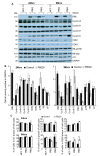
R5020 exerts a biphasic effect on cell cycle regulators and cell cycle progression in MCF-7PRB cells. (A) Protein levels of various cell cycle regulators were analysed by Western Blotting analysis after 24 h and 96 h treatment with R5020. GAPDH was probed as loading control. (B) Densitometry analysis of Western blots in (A) The optical density was quantitated by image J. The results are expressed relative to the intensity of GAPDH for each sample. The data are from two independent experiments with values from PRB2 and PRB3 considered as individual observations, giving rise to n = 4 in each group. The results are expressed as mean ± SEM. GAPDH was used as a loading control for normalization. (C) R5020 enhanced S-phase cells after 24 h but reduced it after 96 h in MCF-7PRB cells. The cell cycle distribution of the individual clones of the MCF-7C and MCF-7PRB cells were determined by flow cytometry analysis of PI staining. Data are presented as the percentage of the cells in the G1, S, and G2/M phase of the cell cycle (mean ± SEM, n = 9 from 3 independent experiments). The degree of statistical significance is indicated with asterisks (* p < 0.05, ** p < 0.01, *** p < 0.001, **** p < 0.001).
Figure 3
Figure 3
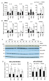
R5020 activates NF-κB pathway and SASP in MCF-7PRB cells. (A) Relative gene expression of IL-1α, IL-1β, IL-8, and NFKBIA at 24 h after R5020 treatment. (B) Relative gene expression of IL-1α, IL-1β, IL-8, and NFKBIA at 96 h after treatment. The data are representative of three independent experiments (mean ± SEM, n = 9). (C) Protein levels of phosphorylated and total IkBα and p65 over a 96-h period were analyzed by Western blotting. GAPDH was probed as a loading control. (D) R5020 treatment increased the ratio of IkBα-pS32/36/IkBα and p65-pS536/p65. The optical density was obtained by image J. The results are normalized to the intensity of GAPDH for each sample. The data are from two independent experiments with values from PRB2 and PRB3 considered as individual observations, giving rise to n = 4 in each group. The results are expressed as mean ± SEM. Asterisks indicate the statistical significance between R5020 against control (* p < 0.05, ** p < 0.01, *** p < 0.001, **** p < 0.001.
Figure 4
Figure 4
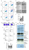
R5020-treated MCF-7PRB cells were unable to form tumorspheres and underwent massive apoptosis. (A) R5020 increased CD24+CD44+ cell population in MCF-7PRB cells. Cells were treated with R5020 for 96 h before being stained with anti-CD44-PE and anti-CD24-FITC antibodies for Flow cytometric analysis. Quadrants were set using appropriate controls. The numbers in Q2 are the average percentage of CD24+CD44+ cells from three independent experiments (Mean ± SEM, n = 9). (B) R5020-treated cells were unable to form tumorspheres. Cells were treated with 10 nM R5020 for 72 h before being plated onto Matrigel for tumorsphere growth, and the images were taken on day 14 after plating. (C) R5020-treated cells did not survive the tumorsphere culture. Cells from B. were released from Matrigel by dispase and trypsin digestion before viable cells were counted after staining with trypan blue. (D) R5020-treated MCF-7PRB cells did not survive when plated in medium containing 2% Matrigel. Cells were treated with 10 nM R5020 for 72 h before being plated in medium containing 2% Matrigel and 10 nM R5020. The cells were cultured in their respective treatment for 14 days before they were stained with 0.1% crystal violet. (E) R5020-treated MCF-7PRB cells underwent apoptosis, as analyzed by a TUNEL assay. MCF-7PRB cells were treated with 10 nM R5020 for 72 h before being plated in 2% Matrigel. After 48 h, DNA fragmentation was evaluated by a FACS-based TUNEL assay. The FITC-A- indicates the percentage of cells with undamaged DNA. FITC-A+ indicates the percentage of cells with fragmented DNA. (F) The percentage of FITC-positive cells are presented as the mean ± SEM, n = 9 from 3 independent experiments. (G) Analysis of the apoptosis-related proteins by Western blotting analysis. Cells were treated with 10 nM R5020 for 72 h before being seeded in 2% Matrigel and cell lysates were collected for Western blotting analysis at 48 h plating. GAPDH was analyzed as loading control. The degree of statistical significance is indicated with asterisks (** p < 0.01, **** p < 0.001).
Figure 5
Figure 5
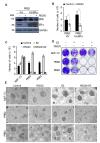
R5020 abolished estrogen-induced cell growth and tumorsphere formation. (A,B) cells were plated in 5% DCC-FCS medium and transduced with adenoviral control vector (Ad) or Ad-ERα. A total of 24 h after the transduction, cells were treated with 10 nM R5020 for 96 h. (A) Western blotting analysis of PR and ERα protein in Ad and Ad-ERα-transduced cells, and GAPDH was analyzed as the loading control. (B) ERα overexpression did not affect R5020-induced growth inhibition in MCF-7PRB cells. (C) R5020 abolished estrogen-induced growth in MCF-7-PRB cells after treatment for 96 h. R5020 treated cells are unable to survive in 2% Matrigel (D) or to form tumorspheres with or without E2 treatment (E). All numeric data are presented as the mean ± SEM, n = 9 from 3 independent experiments. The degree of statistical significance between control and R5020 treatment is indicated by Asterisks (* p < 0.05, ** p < 0.01).
Figure 6
Figure 6
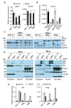
R5020 represses the expression ofERα and FOXA1 and their chromatin association. (A) R5020 treatment of MCF-7PRB cells for 3 h significantly down-regulated the expression of ESR1 and FOXA1. (B) Treatment with cycloheximide (CHX) for 6 h was unable to abolish R5020 repression of FOXA1 gene expression. (C) Treatment with R5020 markedly reduced protein levels of ERα and FOXA1 in MCF-7PRB cells, as determined by Western blotting analysis. GAPDH was probed as a loading control. (D) R5020 diminished chromatin-bound ERα and FOXA1 in a cell fractionation assay. GAPDH, EZH2, and H3 were used as markers for cytoplasmic, nuclear, and chromatin-bound fractions, respectively. (E) R5020 treatment for 24 h abolished estrogen-induced expression of pS2 and AREG in MCF-7PRB cells. All numeric data are presented from three independent experiments (mean ± SEM, n = 9). The degree of statistical significance is indicated with asterisks (* p < 0.05, **** p < 0.001).

References

    1. Osborne C.K., Schiff R. Mechanisms of endocrine resistance in breast cancer. Annu. Rev. Med. 2011;62:233–247. doi: 10.1146/annurev-med-070909-182917. - DOI - PMC - PubMed
    1. Sathyamoorthy N., Lange C.A. Progesterone and Breast Cancer: An NCI Workshop Report. Horm. Cancer. 2020;11:1–12. doi: 10.1007/s12672-020-00379-1. - DOI - PMC - PubMed
    1. Hagan C.R., Lange C.A. Molecular determinants of context-dependent progesterone receptor action in breast cancer. BMC Med. 2014;12:32. doi: 10.1186/1741-7015-12-32. - DOI - PMC - PubMed
    1. Rossouw J.E., Anderson G.L., Prentice R.L., LaCroix A.Z., Kooperberg C., Stefanick M.L., Jackson R.D., Beresford S.A., Howard B.V., Johnson K.C., et al. Risks and benefits of estrogen plus progestin in healthy postmenopausal women: Principal results From the Women’s Health Initiative randomized controlled trial. JAMA. 2002;288:321–333. doi: 10.1001/jama.288.3.321. - DOI - PubMed
    1. Beral V. Breast cancer and hormone-replacement therapy in the Million Women Study. Lancet. 2003;362:419–427. doi: 10.1016/S0140-6736(03)14596-5. - DOI - PubMed