. 2022 Aug 2;10(8):1867.
doi: 10.3390/biomedicines10081867.

Stabilization of F-Actin Cytoskeleton by Paclitaxel Improves the Blastocyst Developmental Competence through P38 MAPK Activity in Porcine Embryos

Affiliations

Stabilization of F-Actin Cytoskeleton by Paclitaxel Improves the Blastocyst Developmental Competence through P38 MAPK Activity in Porcine Embryos

Seung-Yeon Joe et al. Biomedicines. .

Abstract

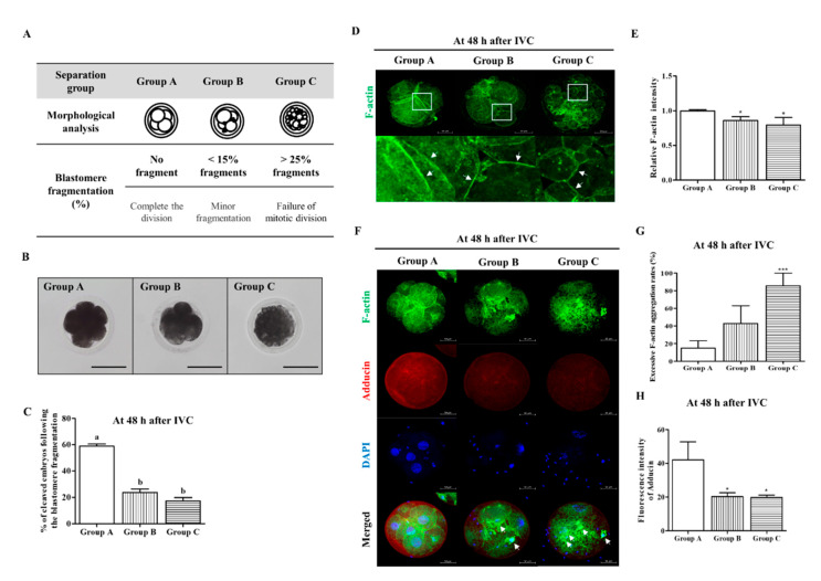
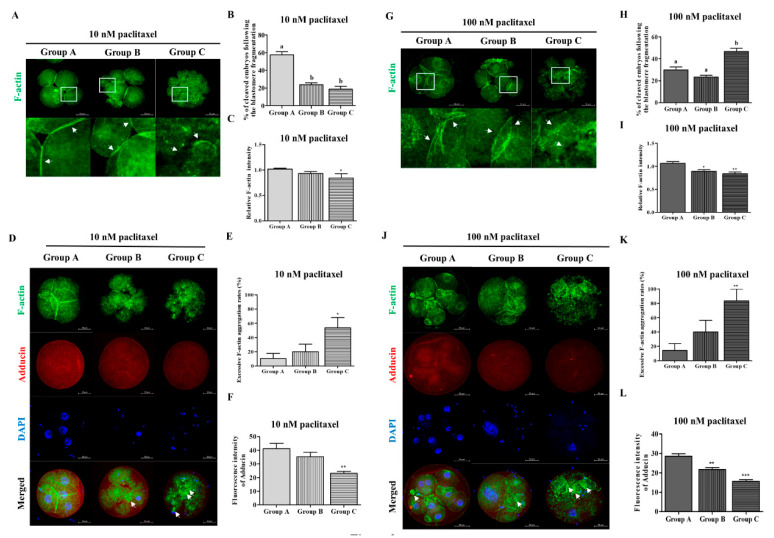
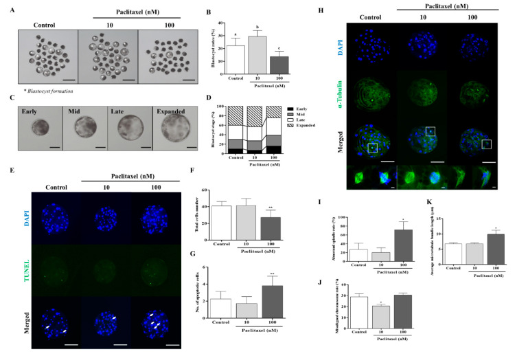
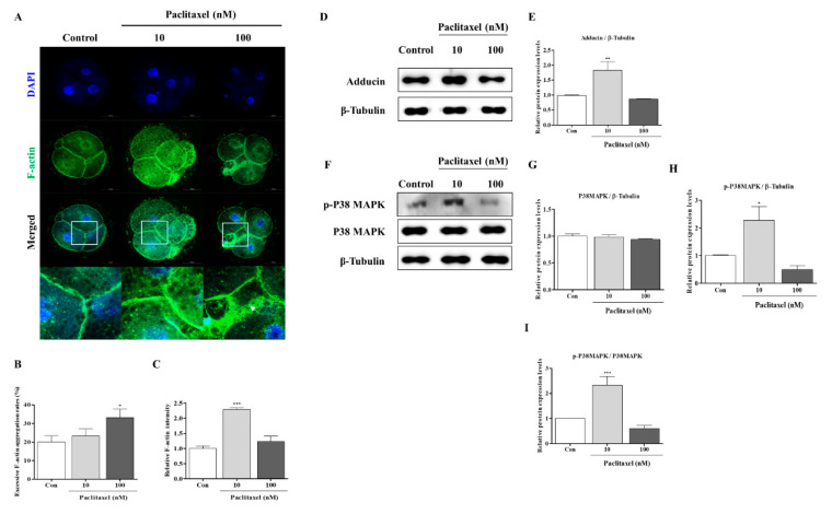
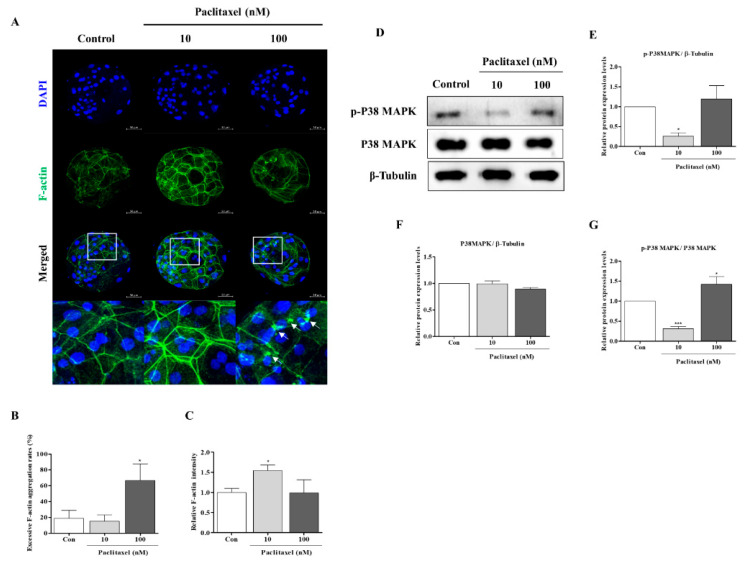
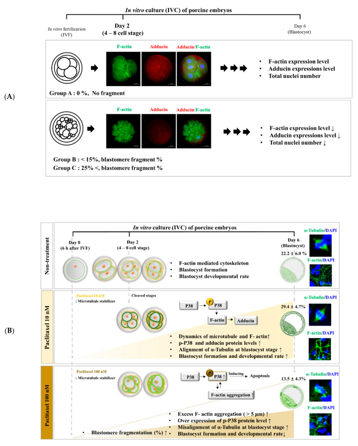
Changes in F-actin distribution and cortical F-actin morphology are important for blastocyst developmental competence during embryogenesis. However, the effect of paclitaxel as a microtubule stabilizer on embryonic development in pigs remains unclear. We investigated the role of F-actin cytoskeleton stabilization via P38 MAPK activation using paclitaxel to improve the developmental potential of blastocysts in pigs. In this study, F-actin enrichment and adducin expression based on blastomere fragment rate and cytokinesis defects were investigated in cleaved embryos after in vitro fertilization (IVF). Adducin and adhesive junction F-actin fluorescence intensity were significantly reduced with increasing blastomere fragment rate in porcine embryos. In addition, porcine embryos were cultured with 10 and 100 nM paclitaxel for two days after IVF. Adhesive junction F-actin stabilization and p-P38 MAPK activity in embryos exposed to 10 nM paclitaxel increased significantly with blastocyst development competence. However, increased F-actin aggregation, cytokinesis defects, and over-expression of p-P38 MAPK protein by 100 nM paclitaxel exposure disrupted blastocyst development in porcine embryos. In addition, exposure to 100 nM paclitaxel increased the misaligned α-tubulin of spindle assembly and adhesive junction F-actin aggregation at the blastocyst stage, which might be caused by p-P38 protein over-expression-derived apoptosis in porcine embryos.

Keywords: F-actin; P38 MAPK; adducin; cytoskeleton; paclitaxel; porcine embryo.


Conflict of interest statement

The authors declare no conflict of interest.

Figures

Figure 1
Figure 1
Porcine embryos reduced accumulation of adhesive junction F-actin and adducin expression according to blastomere fragment rate. (A) Typical images were divided into three groups (Groups A, B, and C) based on morphological criteria of blastomere in porcine embryos at the cleavage stage. Group A: Equal size blastomeres and completed the division state without fragmentation. Group B: Minor fragmentation state with less than 15%. Group C: Unequal size blastomeres with more than 25% fragments (failure of mitotic division state). Scale bar = 150 μm. (B) Bright-field micrograph of porcine embryos (4- or 8-cell stage) in three groups (Groups A, B, and C) according to the presence or absence of blastomere fragmentation. (C) Change in cleavage rate (%) of porcine embryos according to the blastomere fragmentation percentage. The data are expressed as means ± SD. Different superscripts denote a significant difference (p < 0.05). (D,E) In porcine embryos at the cleaved stage, accumulation or intensity of adhesive junction F-actin expression (white arrows) was reduced with increasing blastomere fragment ratio. Scale bar = 50 μm. (FH) Confirmation of changing F-actin aggregation, co-location of F-actin, and adducin fluorescence expression in Groups A, B, and C embryos. Fluorescence expressions were presented with F-actin (green), adducin (red), and nuclei (blue) using immunofluorescence and 4′,6-diamidino-2-phenylindole (DAPI) staining. Scale bar = 50 μm. Data in the bar graph are presented as the mean ± SD of three independent experiments. Differences were considered significant at * p < 0.05 and *** p < 0.001 as compared to the group A.
Figure 2
Figure 2
Accumulation of adhesive junction F-actin, aggregation F-actin and adducin expression by paclitaxel exposure in porcine embryos from Groups A, B, and C. (B,H) Comparison of cleaved embryos ratio from Groups A, B, and C following blastomere fragmentation according to 10 nM (B) and 100 nM (H) paclitaxel exposure in pigs. (A,G) Accumulation of adhesive junction F-actin was observed in the peri-cleavage regions (white arrows) in blastomere of cleaved embryos from Groups A, B, and C. (C,I) Fluorescent expression intensity of F-actin in 10 nM and 100 nM paclitaxel exposed embryos. Data are expressed as means ± SD. Different superscripts denote a significant difference (p < 0.05). (DF,JL) Fluorescence expressions were presented with F-actin (green), adducin (red), and nuclei (blue) using IF and DAPI staining in porcine cleaved embryos from paclitaxel treatment (10 and 100 nM). Scale bar = 50 μm. Data in the bar graph are presented as the mean ± SD of three independent experiments. Differences were considered significant at * p < 0.05, ** p < 0.01, and *** p < 0.001 as compared to the Group A.
Figure 3
Figure 3
Effects of paclitaxel exposure on blastocyst developmental competence and quality in porcine embryos. (A,B) Representative photographs and total blastocyst formation number (white asterisks) by 10 and 100 nM paclitaxel exposure on porcine embryo development. Scale bar = 150 μm. (C,D) Percentage of blastocyst developmental rate as Early, Mid, Late, and Expanded stage after 10 and 100 nM paclitaxel treatment in porcine embryos. Data represent at least three independent experiments and are shown as means ± SD. Different superscript letters denote a significant difference (p < 0.05). (EG) Detection of apoptotic cells and total nuclei in 10 and 100 nM paclitaxel exposed blastocysts using TUNEL (green, white arrow) and DAPI (blue) staining. The graphs show the number of total nuclei and apoptotic embryo rates in blastocysts treated with 10 and 100 nM paclitaxel. Scale bar = 50 μm. (HK) Fluorescence expression of α-tubulin formation related to spindle assembly and chromosome alignment in developed porcine blastocyst after 10 and 100 nM paclitaxel treatment (α-tubulin: green and DAPI: blue). Data are expressed as mean ± SD and were analyzed using one-way ANOVA followed by Tukey’s multiple comparison test. Differences were considered significant at * p < 0.05 and ** p < 0.01.
Figure 4
Figure 4
Changes of adhesive junction F-actin, P38 MAPK, and adducin protein levels in paclitaxel exposed porcine embryos at the cleavage stage. (AC) Confirmation of adhesive junction F-actin expression by diverse paclitaxel exposure (10 and 100 nM) during the cleaved embryo stage in pigs. Fluorescence expression of F-actin (green) and DAPI (blue) in cleaved embryos treated with paclitaxel after IVC. Scale bar = 50 μm. (D,E) The protein level of adducin as an actin-binding protein in 10 and 100 nM paclitaxel exposed embryos. (FI) Western blotting results of the P38 MAPK signal (p-P38 and P38) proteins in 10 and 100 nM paclitaxel treated embryos. Relative folds of protein levels were obtained by normalizing the signals to β-tubulin. Blots were probed with phosphorylation-specific antibodies for P38 MAPK and total P38 MAPK. Densitometric analysis was performed by normalizing phosphorylated P38 MAPK to total levels of P38 MAPK. Histograms represent the values of densitometry analysis obtained using ImageJ software. Data in the bar graph are presented as the means ± SD of three independent experiments (per 70 cleaved embryos). Differences were considered significant at * p < 0.05, ** p < 0.01, *** p < 0.001 compared to the control group.
Figure 5
Figure 5
Confirmation of relationship with adhesive junction F-actin enrichment and P38 MAPK activation in developed porcine blastocysts after 10 and 100 nM paclitaxel exposure. (AC) Changing adhesive junction F-actin expression (white arrows) by paclitaxel exposure (10 and 100 nM) at the blastocyst stage in pigs. Fluorescence expression of F-actin (green) and DAPI (blue) in the blastocyst stage treated with paclitaxel after IVC. Scale bar = 50 μm. Data on the bar graph are presented as the mean ± SD of three independent experiments. Differences were considered significant at * p < 0.05 compared to the control group. (DG) Protein levels of P38 MAPK (p-P38 and P38) using Western blotting in developed porcine blastocyst from 10 and 100 nM paclitaxel treated embryos. Relative folds of protein levels were obtained by normalizing the signals to β-tubulin. Blots were probed with phosphorylation-specific antibodies for P38 MAPK and total P38 MAPK. Densitometric analysis was done by normalizing phosphorylated P38 MAPK to total levels of P38 MAPK. Histograms represent the values of densitometry analysis obtained using ImageJ software. Data in the bar graph are presented as the means ± SD of three independent experiments (per 50 blastocysts). Differences were considered significant at * p < 0.05, *** p < 0.001 compared to the control group.
Figure 6
Figure 6
Graphical summary. Schematic diagram illustrating the correlation between F-actin/adducin cytoskeleton morphology and blastomere fragment rate related to porcine embryo quality during early embryonic development stages. ((A): Top panel); Embryos with blastomere fragment significantly decreased the developmental competence until blastocyst. In addition, Group A embryos showed the improving blastocyst developmental competence by F-actin and adducin related microtubule distribution and dynamics compared with Groups B and C. Simultaneously, low fluorescent expression of F-actin and adducin were observed in embryos from Groups B and C. Therefore, F-actin aggregation and adhesive junction F-actin enrichment can be suggested as a standard for evaluating the quality of embryos due to blastomere division in porcine embryos from IVC. ((B): Bottom panel) Abnormal cleaved blastomeres or cytokinesis defects of embryos are connected to changing F-actin distribution and enrichment at the adhesive junction site in pigs (non-treatment). Interestingly, the 10 nM paclitaxel exposed porcine embryos improve blastocyst developmental capacity through reinforced F-actin filament, adhesive junction F-actin enrichment, and increased adducin protein level in porcine embryos at the cleavage stage. Additionally, 10 nM paclitaxel also induced the p-P38 MAPK activation, improving the firmness of adhesive junction F-actin and microtubule alignment of spindle assembly at the blastocyst stage. However, 100 nM paclitaxel exposed embryos disrupted F-actin distribution, accumulated adhesive junction F-actin, and reduced adducin protein levels in porcine embryos during cleaved stages. The collapse of F-actin enrichments in embryos by 100 nM paclitaxel exposure led to the blastocyst stage with microtubule misalignment, induction of F-actin aggregation, and p-P38 MAPK over-expression. Based on these findings, we propose a positive effect of 10 nM paclitaxel for improving embryo development rate, blastocyst formation and quality via F-actin morphology, aggregation, and enrichment at the adhesive junction site of cleaving embryos in pigs.

Similar articles

Cited by

References

    1. Buey R.M., Barasoain I., Jackson E., Meyer A., Giannakakou P., Paterson I., Mooberry S., Andreu J.M., Díaz J.F. Microtubule Interactions with Chemically Diverse Stabilizing Agents: Thermodynamics of Binding to The Paclitaxel Site Predicts Cytotoxicity. Chem. Biol. 2005;12:1269–1279. doi: 10.1016/j.chembiol.2005.09.010. - DOI - PubMed
    1. Yang C.-P.H., Horwitz S.B. Taxol®: The First Microtubule Stabilizing Agent. Int. J. Mol. Sci. 2017;18:1733. doi: 10.3390/ijms18081733. - DOI - PMC - PubMed
    1. Leung J.C., Cassimeris L. Reorganization of paclitaxel-Stabilized Microtubule Arrays at Mitotic Entry: Roles of Depolymerizing Kinesins and Severing Proteins. Cancer Biol. Ther. 2019;20:1337–1347. doi: 10.1080/15384047.2019.1638678. - DOI - PMC - PubMed
    1. Weaver B.A. How Taxol/paclitaxel kills cancer cells. Mol. Biol. Cell. 2014;25:2677–2681. doi: 10.1091/mbc.e14-04-0916. - DOI - PMC - PubMed
    1. Yue J., López J.M. Understanding MAPK Signaling Pathways in Apoptosis. Int. J. Mol. Sci. 2020;21:2346. doi: 10.3390/ijms21072346. - DOI - PMC - PubMed

LinkOut - more resources