. 2022 Aug 5;10(8):1902.
doi: 10.3390/biomedicines10081902.

Elevated Vascular Sympathetic Neurotransmission and Remodelling Is a Common Feature in a Rat Model of Foetal Programming of Hypertension and SHR

Affiliations

Elevated Vascular Sympathetic Neurotransmission and Remodelling Is a Common Feature in a Rat Model of Foetal Programming of Hypertension and SHR

Maria Sofia Vieira-Rocha et al. Biomedicines. .

Abstract

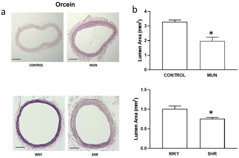
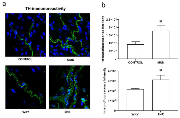
Hypertension is of unknown aetiology, with sympathetic nervous system hyperactivation being one of the possible contributors. Hypertension may have a developmental origin, owing to the exposure to adverse factors during the intrauterine period. Our hypothesis is that sympathetic hyperinnervation may be implicated in hypertension of developmental origins, being this is a common feature with essential hypertension. Two-animal models were used: spontaneously hypertensive rats (SHR-model of essential hypertension) and offspring from dams exposed to undernutrition (MUN-model of developmental hypertension), with their respective controls. In adult males, we assessed systolic blood pressure (SBP), diastolic blood pressure (DBP), heart rate (HR), sympathetic nerve function (3H-tritium release), sympathetic innervation (immunohistochemistry) and vascular remodelling (histology). MUN showed higher SBP/DBP, but not HR, while SHR exhibited higher SBP/DBP/HR. Regarding the mesenteric arteries, MUN and SHR showed reduced lumen, increased media and adventitial thickness and increased wall/lumen and connective tissue compared to respective controls. Regarding sympathetic nerve activation, MUN and SHR showed higher tritium release compared to controls. Total tritium tissue/tyrosine hydroxylase detection was higher in SHR and MUN adventitia arteries compared to respective controls. In conclusion, sympathetic hyperinnervation may be one of the contributors to vascular remodelling and hypertension in rats exposed to undernutrition during intrauterine life, which is a common feature with spontaneous hypertension.

Keywords: fibrosis; foetal programming of hypertension; foetal undernutrition; sympathetic innervation; sympathetic neurotransmission; vascular remodelling.


Conflict of interest statement

The authors declare no conflict of interest.

Figures

Figure 1
Figure 1
Histomorphometry of the lumen of mesenteric arteries from CONTROL and MUN (upper panel) and WKY and SHR rats (lower panel). (a) Images were obtained from orcein stained arteries (scale bar = 500 µm); (b) the graphics show the lumen area. MUN, offspring exposed to maternal undernutrition during pregnancy; CONTROL, offspring from mothers fed ad libitum during pregnancy; SHR, spontaneously hypertensive rats; WKY, Wistar Kyoto rats. Values are mean ± s.e.m. from 6 rats of each group. Significant differences from the respective control rat: * p < 0.05.
Figure 2
Figure 2
Histomorphometry of hypertensive (MUN and SHR) and normotensive (CONTROL and WKY) mesenteric wall. (a) Images were obtained from haematoxylin/eosin-stained arteries (scare bar = 300 µm). (b) Cross-sectional area of tunica media (right panel) and cross-sectional area of tunica adventitia (left panel). MUN, offspring exposed to maternal undernutrition during pregnancy; CONTROL, offspring from mothers fed ad libitum during pregnancy; SHR, spontaneously hypertensive rats; WKY, Wistar Kyoto rats. Values are mean ± s.e.m. from 6 rats of each group. Significant differences from the respective control rat: * p < 0.05; ** p < 0.01 *** p < 0.001.
Figure 3
Figure 3
Histomorphometry of hypertensive (MUN and SHR) and normotensive (CONTROL and WKY) mesenteric arteries, stained with Trichrome. MUN, offspring exposed to maternal undernutrition during pregnancy; CONTROL, offspring from mothers fed ad libitum during pregnancy; SHR, spontaneously hypertensive rats; WKY, Wistar Kyoto rats. Values are mean ± s.e.m. from 6 rats of each group. Significant differences from the respective control rat: * p < 0.05.
Figure 4
Figure 4
Representative examples of time course tritium outflow from: mesenteric arteries from normotensive animals, CONTROL (circles, left panel) and WKY (circles, right panel), and hypertensive animals, MUN (triangles, left panel) and SHR (triangles, right panel) from typical experiments. After pre-incubation with [3H]-noradrenaline, tissues were superfused with [3H]-noradrenaline-free medium containing desipramine (400 nM). Tritium outflow (ordinates) is expressed as a percentage of the total radioactivity present in the tissue at the beginning of the collection period and was measured in samples collected every 5 min. Artery segments were stimulated twice by 100 pulses/5 Hz, (S1, S2). Each line represents the outflow of tritium from a single superfusion chamber; MUN, offspring exposed to maternal undernutrition during pregnancy; CONTROL, offspring from mothers fed ad libitum during pregnancy; SHR, spontaneously hypertensive rats; WKY, Wistar Kyoto rats.
Figure 5
Figure 5
[3H]-Tritium uptake in mesenteric arteries from (a) CONTROL and MUN and (b) WKY and SHR rats. MUN, offspring exposed to maternal undernutrition during pregnancy; CONTROL, offspring from mothers fed ad libitum during pregnancy; SHR, spontaneously hypertensive rats; WKY, Wistar Kyoto rats. Values are mean ± s.e.m. from 6 animals per group. Significant differences from the respective control rats: * p < 0.05.
Figure 6
Figure 6
(a) Laser scanning confocal microscopy representative images of the adventitia layer of mesenteric arteries from CONTROL and MUN (upper panel) WKY and SHR (lower panel) rats. Images show the immunofluorescence reactivity to TH (green) with DAPI-stained nuclei (blue). Scale bar: 25 μm. (b) Quantitative analysis of LSCM images. MUN, offspring exposed to maternal undernutrition during pregnancy; CONTROL, offspring from mothers fed ad libitum during pregnancy; SHR, spontaneously hypertensive rats; WKY, Wistar Kyoto rats. In the graphics, values are mean ± s.e.m. from 6 rats from each group. Significant differences from the respective control rats: * p < 0.05.
Figure 7
Figure 7
Effect of suboptimal foetal undernutrition on SNS and vascular wall from a rat FPH model.

Similar articles

Cited by

References

    1. van der Linden E.L., Collard D., Beune E.J.A.J., Nieuwkerk P.T., Galenkamp H., Haafkens J.A., van Charante E.P.M., van den Born B.-J.H., Agyemang C. Determinants of suboptimal blood pressure control in a multi-ethnic population: The Healthy Life in an Urban Setting (HELIUS) study. J. Clin. Hypertens. 2021;23:1068–1076. doi: 10.1111/jch.14202. - DOI - PMC - PubMed
    1. Thomas P., Dasgupta I. The role of the kidney and the sympathetic nervous system in hypertension. Pediatr. Nephrol. 2015;30:549–560. doi: 10.1007/s00467-014-2789-4. - DOI - PubMed
    1. Oparil S., Zaman M.A., Calhoun D.A. Pathogenesis of hypertension. Ann. Inter. Med. 2003;139:761–776. doi: 10.7326/0003-4819-139-9-200311040-00011. - DOI - PubMed
    1. Grassi G., Bertoli S., Seravalle G. Sympathetic nervous system: Role in hypertension and in chronic kidney disease. Curr. Opin. Nephrol. Hypertens. 2012;21:46–51. doi: 10.1097/MNH.0b013e32834db45d. - DOI - PubMed
    1. Grassi G., Ram V.S. Evidence for a critical role of the sympathetic nervous system in hypertension. J. Am. Soc. Hypertens. 2016;10:457–466. doi: 10.1016/j.jash.2016.02.015. - DOI - PubMed

LinkOut - more resources