Review
. 2022 Aug 6;10(8):1906.
doi: 10.3390/biomedicines10081906.

The Journey of Human Transthyretin: Synthesis, Structure Stability, and Catabolism

Affiliations
Review

The Journey of Human Transthyretin: Synthesis, Structure Stability, and Catabolism

Chiara Sanguinetti et al. Biomedicines. .

Abstract

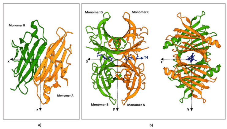
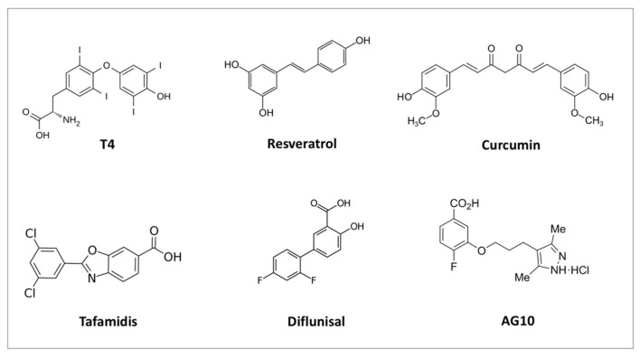
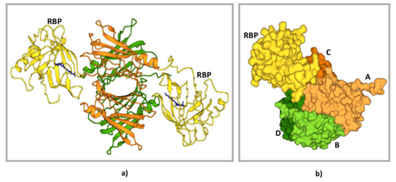
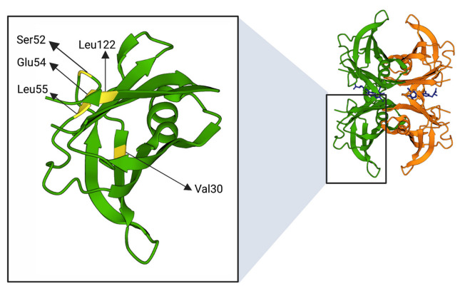
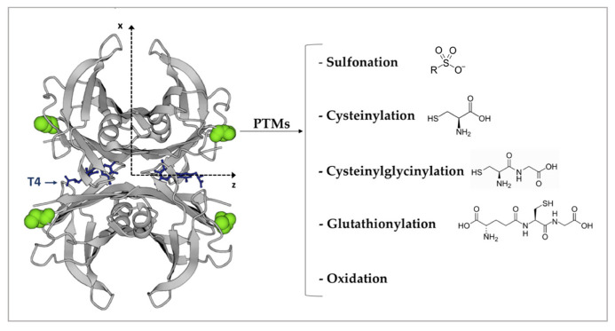
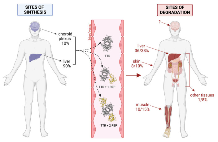
Transthyretin (TTR) is a homotetrameric protein mainly synthesised by the liver and the choroid plexus whose function is to carry the thyroid hormone thyroxine and the retinol-binding protein bound to retinol in plasma and cerebrospinal fluid. When the stability of the tetrameric structure is lost, it breaks down, paving the way for the aggregation of TTR monomers into insoluble fibrils leading to transthyretin (ATTR) amyloidosis, a progressive disorder mainly affecting the heart and nervous system. Several TTR gene mutations have been characterised as destabilisers of TTR structure and are associated with hereditary forms of ATTR amyloidosis. The reason why also the wild-type TTR is intrinsically amyloidogenic in some subjects is largely unknown. The aim of the review is to give an overview of the TTR biological life cycle which is largely unknown. For this purpose, the current knowledge on TTR physiological metabolism, from its synthesis to its catabolism, is described. Furthermore, a large section of the review is dedicated to examining in depth the role of mutations and physiological ligands on the stability of TTR tetramers.

Keywords: ER-associated degradation pathway; TTR amyloidosis; TTR clearance; retinol; retinol-binding protein; thyroxine; transthyretin.


Conflict of interest statement

The authors declare no conflict of interest.

Figures

Figure 1
Figure 1
TTR structure. (a) Ribbon diagram of the AB dimer interface (b) Ribbon diagram of the TTR tetramers. The X-axis passes through the T4 binding channels, which are formed at the interface of monomers D-B and C-A. The halogen binding pockets (HBP) are shaped by the following amino acids: HBP1, Lys15-Leu17-Thr106-Val 121; HBP2, Lys15-Leu17-Ala108-Ala109-Leu110; HBP3, Ser117-Leu110-Thr119-Ala108. T4 is represented in blue. The figure has been produced using “www.rcsb.org”web site (protein ID code 1QAB), access date 22 June 2022.
Figure 2
Figure 2
Chemical structure of some natural (T4, resveratrol, curcumin) and chemical ligands (tafamidis, diflunisal, AG10) able to bind and stabilise TTR tetramer.
Figure 3
Figure 3
Structure of TTR–RBP complex. (a) Structure representation of the TTR–RBP complex. TTR: monomers A and C, orange; monomers B and D, green. RBP: yellow. Retinol: blue (b) Detail of the contact between the TTR subunits A, C, and D and the RBP molecule [left side, colour codes as in (a)]. Centre and right drawings show the interacting surfaces of RBP (centre) and of the TTR subunits A, C, and D (right). It is possible to appreciate how the RBP surface fits into a site formed by the arrangement of three TTR subunits (A, C, and D). The figure has been produced using “www.rcsb.org” web site, access date 22 June 2022.
Figure 4
Figure 4
Positions of amino acids (highlighted in yellow) subjected to mutations described in Table 1. The figure was created through “www.rcsb.org” and “biorender.com” web sites (license agreement number KJ247URGZS), access date 22 June 2022.
Figure 5
Figure 5
The four Cys 10 are highlighted in green and represented as a spacefill model. The most frequent Cys10 PTMs are indicated on the right. T4 is represented in blue. The figure has been produced by using “www.rcsb.org” web site (protein ID code 1QAB), access date 22 June 2022.
Figure 6
Figure 6
Reconstruction of the possible life cycle of TTR. The figure was created through “biorender.com” web site (license agreement number DL2489YBZI), access date 22 June 2022.

References

    1. Robbins J. Transthyretin from discovery to now. Clin. Chem. Lab. Med. (CCLM) 2002;40:1183–1190. doi: 10.1515/CCLM.2002.208. - DOI - PubMed
    1. Nomenclature committee of the International Union of Biochemistry (NC-IUB) Enzyme nomenclature. Recommendations 1978. Supplement 2: Corrections and additions. Eur. J. Biochem. 1981;116:423–435. doi: 10.1111/j.1432-1033.1981.tb05353.x. - DOI - PubMed
    1. Noy N., Slosberg E., Scarlata S. Interactions of retinol with binding proteins: Studies with retinol-binding protein and with transthyretin. Biochemistry. 1992;31:11118–11124. doi: 10.1021/bi00160a023. - DOI - PubMed
    1. Nilsson S., Rask L., Peterson P. Studies on thyroid hormone-binding proteins. II. Binding of thyroid hormones, retinol-binding protein, and fluorescent probes to prealbumin and effects of thyroxine on prealbumin subunit self association. J. Biol. Chem. 1975;250:8554–8563. doi: 10.1016/S0021-9258(19)40795-3. - DOI - PubMed
    1. Wei S., Episkopou V., Piantedosi R., Maeda S., Shimada K., Gottesman M.E., Blaner W.S. Studies on the metabolism of retinol and retinol-binding protein in transthyretin-deficient mice produced by homologous recombination. J. Biol. Chem. 1995;270:866–870. doi: 10.1074/jbc.270.2.866. - DOI - PubMed