Review
. 2022 Aug 11;10(8):1943.
doi: 10.3390/biomedicines10081943.

Lipid Metabolism in Glioblastoma: From De Novo Synthesis to Storage

Affiliations
Review

Lipid Metabolism in Glioblastoma: From De Novo Synthesis to Storage

Yongjun Kou et al. Biomedicines. .

Abstract

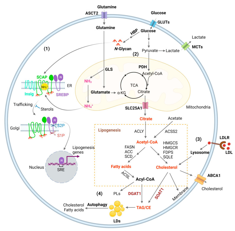
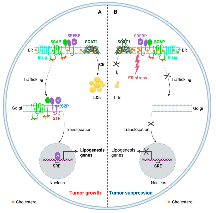
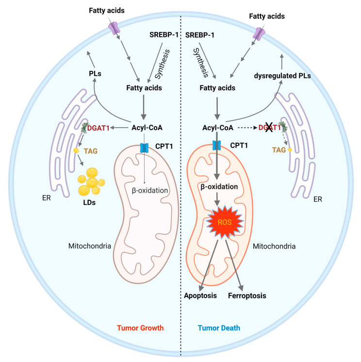
Glioblastoma (GBM) is the most lethal primary brain tumor. With limited therapeutic options, novel therapies are desperately needed. Recent studies have shown that GBM acquires large amounts of lipids for rapid growth through activation of sterol regulatory element-binding protein 1 (SREBP-1), a master transcription factor that regulates fatty acid and cholesterol synthesis, and cholesterol uptake. Interestingly, GBM cells divert substantial quantities of lipids into lipid droplets (LDs), a specific storage organelle for neutral lipids, to prevent lipotoxicity by increasing the expression of diacylglycerol acyltransferase 1 (DGAT1) and sterol-O-acyltransferase 1 (SOAT1), which convert excess fatty acids and cholesterol to triacylglycerol and cholesteryl esters, respectively. In this review, we will summarize recent progress on our understanding of lipid metabolism regulation in GBM to promote tumor growth and discuss novel strategies to specifically induce lipotoxicity to tumor cells through disrupting lipid storage, a promising new avenue for treating GBM.

Keywords: DGAT1; SOAT1; SREBP-1; cholesterol; fatty acids; glioblastoma; lipid droplets; lipotoxicity.


Conflict of interest statement

The authors declare no conflict of interest.

Figures

Figure 1
Figure 1
Lipid metabolism reprogramming in cancer cells. (1) Combined elevation of glucose and glutamine consumption promotes lipogenesis by activating the SREBP/SCAP pathway. Glucose produces N-glycans through HBP (Hexosamine Biosynthesis Pathway), which stabilizes SCAP (SREBP-cleavage activating protein) through N-linked glycosylation of its luminal loops (1 and 7). Glutamine enters the mitochondria and ammonia (NH3) is released by GLS (glutaminase). NH3 is protonated and converted to NH4+, which directly binds to the aspartate D428 and Serine S326/330 in the core of the SCAP transmembrane domains, forming stable hydrogen bonds. This binding triggers a dramatic conformation change in SCAP, leading to its dissociation from Insig (insulin-induced gene), an endoplasmic reticulum (ER)-resident protein. Subsequently, SCAP escorts SREBP (sterol regulatory element-binding protein) to the Golgi, where it is cleaved by two enzymes S1P (site 1 protease) and S2P (site 2 protease) to release its active N-terminal fragment. Finally, the N-terminal domain goes into the nucleus, binds to the SRE (sterol regulatory element) motif located in the promoters of gene involved in lipogenesis to activate their transcription and promote de novo lipid synthesis and tumor growth. (2) Glucose is the main source for lipid synthesis. Glucose via glycolysis breaks down into pyruvate, which enters the mitochondria and is converted to acetyl-CoA by PDH (pyruvate dehydrogenase), followed by condensation with OAA (oxaloacetate) to form citrate to enter the TCA cycle (tricarboxylic acid cycle). Citrate is released to the cytosol via its mitochondria transporter, SLC25A1. Citrate is then cleaved to acetyl-CoA by ACLY (ATP citrate lyase), which serves as a precursor for fatty acid and cholesterol biosynthesis catalyzed by a series of enzymes that are the main transcriptional targets of SREBPs, as shown in the Figure. In addition, cytosol acetate can be converted to acetyl-CoA for lipid synthesis by ACCS2 (acetyl-CoA synthetase 2). Besides that, acetyl-CoA is also the substrate for the acetylation of histones, which is involved in epigenetic regulation. Moreover, glutamine through glutaminolysis contributes as an anaplerotic substrate to replenish tricarboxylic acid (TCA) cycle intermediates. Glutamate, the product of the first step of glutaminolysis, is converted to α-KG (α-ketoglutaric acid) and then enters into the TCA cycle. (3) SREBPs upregulates the expression of LDLR (low-density lipoprotein receptor), which binds to LDL and transports it into cells through the endocytosis process. LDL is then hydrolyzed in the lysosomes and cholesterol is released, promoting tumor growth. (4) Excess fatty acids and cholesterol are converted to TAG (triacylglycerol) and CE (cholesteryl ester) by DGAT1 (diacylglycerol O-acyltransferase 1) and sterol O acyl-transferase 1 (SOAT1) to form LDs (lipid droplets) and prevent toxicity from high lipid levels. Under conditions of nutrient deficiency, LDs are hydrolyzed by autophagy to release free fatty acids and cholesterol for tumor survival.
Figure 2
Figure 2
Targeting SOAT1 to disrupt cholesterol homeostasis for GBM therapy. (A) SOAT1 catalyzes the esterification of cholesterol to store excess free cholesterol into LDs, and thus is involved in maintaining cellular cholesterol homeostasis. Cholesterol negatively regulates SCAP/SREBP trafficking and SREBP activation. Cholesterol reduction in the ER activates SCAP dissociation from Insig, resulting in SREBP translocation and activation to promote lipogenesis. (B) Inhibition of SOAT1 reduces CE formation, which in turn results in the accumulation of cholesterol in the ER membrane, subsequently leading to the SCAP/SREBP complex remaining in the ER and to reduction of lipogenesis, along with enhanced ER stress, resulting in tumor suppression.
Figure 3
Figure 3
Inhibition of DGAT1 as a therapy strategy to induce cancer cell death. (1) Left: Tumor cells acquire fatty acids through uptake or de novo synthesis via the activation of SREBP-1. Fatty acids are converted to acyl-CoAs, which are the substrates for phospholipid (PL) synthesis and produce energy via entering into mitochondria to undergo β-oxidation and oxidative phosphorylation. Excessive acyl-CoA is stored in LDs as catalyzed by DGAT1 in tumor cells. (2) Right: Inhibiting DGAT1 causes an imbalance of fatty acid catabolism, leading to cell death. Inhibiting DGAT1 forces more acyl-CoAs to enter the mitochondria through carnitine palmitate transferase 1 (CPT1) for β-oxidation, leading to high levels of reactive oxygen species (ROS), which trigger apoptosis and ferroptosis to kill tumor cells. Moreover, the profile of PLs is altered upon DGAT1 inhibition, contributing to further cellular stress.

References

    1. Cheng C., Geng F., Cheng X., Guo D. Lipid metabolism reprogramming and its potential targets in cancer. Cancer Commun. 2018;38:27. doi: 10.1186/s40880-018-0301-4. - DOI - PMC - PubMed
    1. Cockcroft S. Mammalian lipids: Structure, synthesis and function. Essays Biochem. 2021;65:813–845. doi: 10.1042/EBC20200067. - DOI - PMC - PubMed
    1. Cisa-Wieczorek S., Hernandez-Alvarez M.I. Deregulation of Lipid Homeostasis: A Fa(c)t in the Development of Metabolic Diseases. Cells. 2020;9:2605. doi: 10.3390/cells9122605. - DOI - PMC - PubMed
    1. Onal G., Kutlu O., Gozuacik D., Dokmeci Emre S. Lipid Droplets in Health and Disease. Lipids Health Dis. 2017;16:128. doi: 10.1186/s12944-017-0521-7. - DOI - PMC - PubMed
    1. Estes R.E., Lin B., Khera A., Davis M.Y. Lipid Metabolism Influence on Neurodegenerative Disease Progression: Is the Vehicle as Important as the Cargo? Front. Mol. Neurosci. 2021;14:788695. doi: 10.3389/fnmol.2021.788695. - DOI - PMC - PubMed