The Dose-Dependent Effects of Multifunctional Enkephalin Analogs on the Protein Composition of Rat Spleen Lymphocytes, Cortex, and Hippocampus; Comparison with Changes Induced by Morphine
- PMID: 36009516
- PMCID: PMC9406115
- DOI: 10.3390/biomedicines10081969
The Dose-Dependent Effects of Multifunctional Enkephalin Analogs on the Protein Composition of Rat Spleen Lymphocytes, Cortex, and Hippocampus; Comparison with Changes Induced by Morphine
Abstract
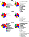
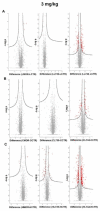
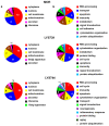
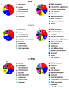
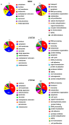
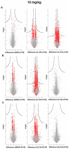
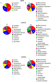
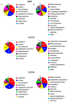
This work aimed to test the effect of 7-day exposure of rats to multifunctional enkephalin analogs LYS739 and LYS744 at doses of 3 mg/kg and 10 mg/kg on the protein composition of rat spleen lymphocytes, brain cortex, and hippocampus. Alterations of proteome induced by LYS739 and LYS744 were compared with those elicited by morphine. The changes in rat proteome profiles were analyzed by label-free quantification (MaxLFQ). Proteomic analysis indicated that the treatment with 3 mg/kg of LYS744 caused significant alterations in protein expression levels in spleen lymphocytes (45), rat brain cortex (31), and hippocampus (42). The identified proteins were primarily involved in RNA processing and the regulation of cytoskeletal dynamics. In spleen lymphocytes, the administration of the higher 10 mg/kg dose of both enkephalin analogs caused major, extensive modifications in protein expression levels: LYS739 (119) and LYS744 (182). Among these changes, the number of proteins associated with immune responses and apoptotic processes was increased. LYS739 treatment resulted in the highest number of alterations in the rat brain cortex (152) and hippocampus (45). The altered proteins were functionally related to the regulation of transcription and cytoskeletal reorganization, which plays an essential role in neuronal plasticity. Administration with LYS744 did not increase the number of altered proteins in the brain cortex (26) and hippocampus (26). Our findings demonstrate that the effect of κ-OR full antagonism of LYS744 is opposite in the central nervous system and the peripheral region (spleen lymphocytes).
Keywords: chronic pain treatment; label-free quantification; morphine; multifunctional enkephalin analogs; proteomic analysis; rat brain; rat spleen lymphocytes.
Conflict of interest statement
The authors declare no conflict of interest.
Figures


Similar articles
-
The impact of multifunctional enkephalin analogs and morphine on the protein changes in crude membrane fractions isolated from the rat brain cortex and hippocampus.Peptides. 2024 Apr;174:171165. doi: 10.1016/j.peptides.2024.171165. Epub 2024 Feb 1. Peptides. 2024. PMID: 38307418
-
The high-resolution proteomic analysis of protein composition of rat spleen lymphocytes stimulated by Concanavalin A; a comparison with morphine-treated cells.J Neuroimmunol. 2020 Apr 15;341:577191. doi: 10.1016/j.jneuroim.2020.577191. Epub 2020 Feb 13. J Neuroimmunol. 2020. PMID: 32113006
-
Impact of three-month morphine withdrawal on rat brain cortex, hippocampus, striatum and cerebellum: proteomic and phosphoproteomic studies.Neurochem Int. 2021 Mar;144:104975. doi: 10.1016/j.neuint.2021.104975. Epub 2021 Jan 27. Neurochem Int. 2021. PMID: 33508371
-
Reflections on: "A general role for adaptations in G-Proteins and the cyclic AMP system in mediating the chronic actions of morphine and cocaine on neuronal function".Brain Res. 2016 Aug 15;1645:71-4. doi: 10.1016/j.brainres.2015.12.039. Epub 2015 Dec 29. Brain Res. 2016. PMID: 26740398 Free PMC article. Review.
-
Role of oxytocin in the neuroadaptation to drugs of abuse.Psychoneuroendocrinology. 1994;19(1):85-117. doi: 10.1016/0306-4530(94)90062-0. Psychoneuroendocrinology. 1994. PMID: 9210215 Review.
References
-
- Bourova L., Vosahlikova M., Kagan D., Dlouha K., Novotny J., Svoboda P. Long-term adaptation to high doses of morphine causes desensitization of µ-OR- and δ-OR-stimulated G-protein response in forebrain cortex but does not decrease the amount of G-protein alpha subunit. Med. Sci. Monit. 2010;16:260–270. - PubMed
-
- Ujcikova H., Dlouha K., Roubalova L., Vosahlikova M., Kagan D., Svoboda P. Up-regulation of adenylylcyclases I and II induced by long-term adaptation of rats to morphine fades away 20 days after morphine withdrawal. Biochim. Biophys. Acta. 2011;1810:1220–1229. doi: 10.1016/j.bbagen.2011.09.017. - DOI - PubMed
Grants and funding
LinkOut - more resources
Full Text Sources

