. 2022 Aug 17;10(8):1991.
doi: 10.3390/biomedicines10081991.

Autologous Platelet-Rich Growth Factor Reduces M1 Macrophages and Modulates Inflammatory Microenvironments to Promote Sciatic Nerve Regeneration

Affiliations

Autologous Platelet-Rich Growth Factor Reduces M1 Macrophages and Modulates Inflammatory Microenvironments to Promote Sciatic Nerve Regeneration

Anjali Yadav et al. Biomedicines. .

Abstract

The failure of peripheral nerve regeneration is often associated with the inability to generate a permissive molecular and cellular microenvironment for nerve repair. Autologous therapies, such as platelet-rich plasma (PRP) or its derivative platelet-rich growth factors (PRGF), may improve peripheral nerve regeneration via unknown mechanistic roles and actions in macrophage polarization. In the current study, we hypothesize that excessive and prolonged inflammation might result in the failure of pro-inflammatory M1 macrophage transit to anti-inflammatory M2 macrophages in large nerve defects. PRGF was used in vitro at the time the unpolarized macrophages (M0) macrophages were induced to M1 macrophages to observe if PRGF altered the secretion of cytokines and resulted in a phenotypic change. PRGF was also employed in the nerve conduit of a rat sciatic nerve transection model to identify alterations in macrophages that might influence excessive inflammation and nerve regeneration. PRGF administration reduced the mRNA expression of tumor necrosis factor-α (TNFα), interleukin-1β (IL-1β), and IL-6 in M0 macrophages. Increased CD206 substantiated the shift of pro-inflammatory cytokines to the M2 regenerative macrophage. Administration of PRGF in the nerve conduit after rat sciatic nerve transection promoted nerve regeneration by improving nerve gross morphology and its targeted gastrocnemius muscle mass. The regenerative markers were increased for regrown axons (protein gene product, PGP9.5), Schwann cells (S100β), and myelin basic protein (MBP) after 6 weeks of injury. The decreased expression of TNFα, IL-1β, IL-6, and CD68+ M1 macrophages indicated that the inflammatory microenvironments were reduced in the PRGF-treated nerve tissue. The increase in RECA-positive cells suggested the PRGF also promoted angiogenesis during nerve regeneration. Taken together, these results indicate the potential role and clinical implication of autologous PRGF in regulating inflammatory microenvironments via macrophage polarization after nerve transection.

Keywords: macrophage polarization; peripheral nerve regeneration; platelet-rich growth factors; sciatic nerve injury.


Conflict of interest statement

The authors declare no conflict of interest.

Figures

Figure 1
Figure 1
PRGF reduces both the secretion and expression of pro-inflammatory cytokines from M1 macrophages. (A) TNFα secretion decreased in M1 macrophages when treated with 5 and 10% PRGF compared to untreated M1 macrophages as checked by ELISA. (B) The concentration of anti-inflammatory cytokine IL-10 increased in M1 macrophages after treatment with 10% PRGF for 24 h. (C) The increases in mRNA expression levels of TNFα, IL-1β, and IL-6 in M1-induction media were inhibited by adding 10% PRGF as checked by qRT-PCR. n = 3. Significance was assessed by one-way ANOVA. Data are presented as the mean ± SEM. * p < 0.05 versus M0. # p < 0.05 versus M1.
Figure 2
Figure 2
PRGF enhanced M2-macrophage polarization and inhibited the LPS-induced inflammatory SCs. (A) Representative phase images revealed a change in cell morphology of PRGF-treated M1 macrophages compared with M1 macrophages. (B) The qRT-PCR showed increases in the mRNA expression level for CD206 and CD86 by treating 10% PRGF with the M1-induction media. (C) The LPS-induced TNFα, IL-1β, and IL-6 expressions were inhibited when mixing with 10% PRGF in RT4 SCs. n = 4. Significance was assessed by one-way ANOVA. Data are presented as the mean ± SEM. For panel (B) histograms; * p < 0.05 versus M0. # p < 0.05 versus M1. For panel (C) histograms; * p < 0.05 versus no LPS and PRGF. # p < 0.05 versus only LPS. Scale bar = 40 μm.
Figure 3
Figure 3
PRGF reduces chronic inflammation and macrophages in nerve conduit. (A) Representative pictures of the harvested sciatic nerves after 6 weeks of transection injury and nerve conduit surgery. (B) Representative immunohistochemistry (IHC) staining images demonstrated the expression levels of TNFα, IL-1β, and IL-6 in the middle region of transverse sectioned nerve tissue were significantly decreased in the rats that received nerve conduit filled with PRGF. (C) The number of M1 macrophages as indicated by the positive marker of CD68 was reduced in PRGF-treated nerve tissue. Scale bar = 20 μm. N = 3. Significance was assessed by paired t-test. Data are presented as the mean ± SEM. * p < 0.05 versus to saline group.
Figure 4
Figure 4
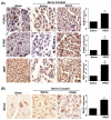
Improvements of nerve regeneration, remyelination, and angiogenesis by PRGF after sciatic nerve injury. (A) The IHC staining for PGP9.5, S100β, and MBP demonstrated increases in axon regrowth, SCs numbers, and remyelination after treatment with PRGF for 6 weeks. (B) An increase in angiogenesis and vascular formation was observed by IHC staining of rat endothelial cell antigen (RECA). Scale bar = 20 μm. N = 3. Significance was assessed by paired t-test. Data are presented as the mean ± SEM. * p < 0.05 versus the saline group.
Figure 5
Figure 5
Myelin sheath formation and target muscle innervation confirmed the regenerative outcomes at 6 weeks post-sciatic nerve injury. (A) Luxol fast blue (LFB) staining showed an increase in myelin sheath formation for the nerve sections that received PRGF treatment as compared to the saline group. (B) The morphology of sciatic nerve innervated target gastrocnemius muscle on an injured (left) and normal (right) leg demonstrated the improvement of regenerated nerve to reinnervate the target muscle for preventing muscle atrophy. (C) The relative gastrocnemius muscle weight (RGMW) was quantified and showed an improvement in the PRGF-treated rats. Scale bar = 20 μm. N = 3. Significance was assessed by paired t-test. Data are presented as the mean ± SEM. * p < 0.05 versus to saline group.

References

    1. Zigmond R.E., Echevarria F.D. Macrophage biology in the peripheral nervous system after injury. Prog. Neurobiol. 2019;173:102–121. doi: 10.1016/j.pneurobio.2018.12.001. - DOI - PMC - PubMed
    1. Msheik Z., El Massry M., Rovini A., Billet F., Desmouliere A. The macrophage: A key player in the pathophysiology of peripheral neuropathies. J. Neuroinflamm. 2022;19:97. doi: 10.1186/s12974-022-02454-6. - DOI - PMC - PubMed
    1. Fregnan F., Muratori L., Simoes A.R., Giacobini-Robecchi M.G., Raimondo S. Role of inflammatory cytokines in peripheral nerve injury. Neural Regen. Res. 2012;7:2259–2266. doi: 10.3969/j.issn.1673-5374.2012.29.003. - DOI - PMC - PubMed
    1. Gaudet A.D., Popovich P.G., Ramer M.S. Wallerian degeneration: Gaining perspective on inflammatory events after peripheral nerve injury. J. Neuroinflamm. 2011;8:110. doi: 10.1186/1742-2094-8-110. - DOI - PMC - PubMed
    1. Liu P., Peng J., Han G.H., Ding X., Wei S., Gao G., Huang K., Chang F., Wang Y. Role of macrophages in peripheral nerve injury and repair. Neural Regen. Res. 2019;14:1335–1342. doi: 10.4103/1673-5374.253510. - DOI - PMC - PubMed