Review
. 2022 Aug 19;10(8):2018.
doi: 10.3390/biomedicines10082018.

Chemerin Forms: Their Generation and Activity

Affiliations
Review

Chemerin Forms: Their Generation and Activity

Lei Zhao et al. Biomedicines. .

Abstract

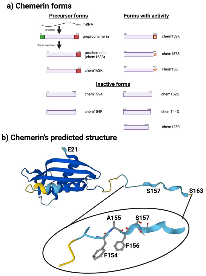
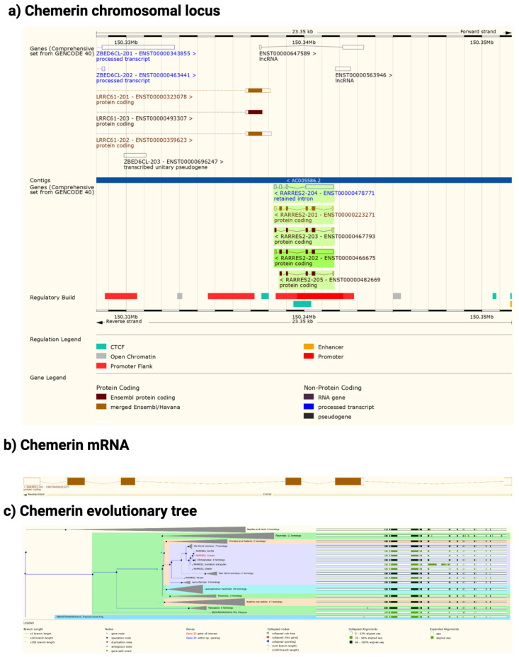
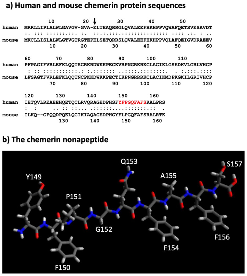
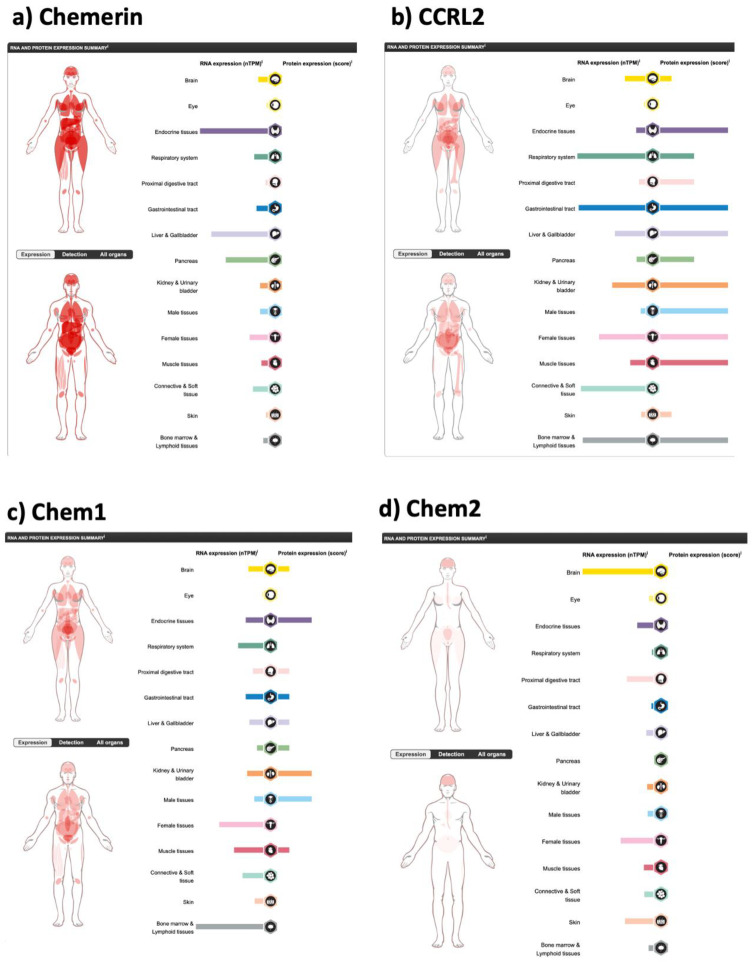
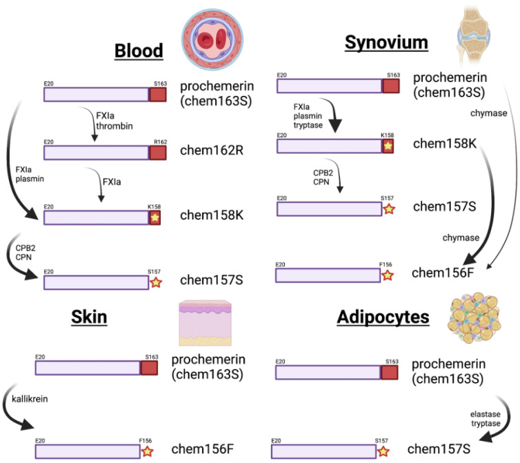
Chemerin is the product of the RARRES2 gene which is secreted as a precursor of 143 amino acids. That precursor is inactive, but proteases from the coagulation and fibrinolytic cascades, as well as from inflammatory reactions, process the C-terminus of chemerin to first activate it and then subsequently inactivate it. Chemerin can signal via two G protein-coupled receptors, chem1 and chem2, as well as be bound to a third non-signaling receptor, CCRL2. Chemerin is produced by the liver and secreted into the circulation as a precursor, but it is also expressed in some tissues where it can be activated locally. This review discusses the specific tissue expression of the components of the chemerin system, and the role of different proteases in regulating the activation and inactivation of chemerin. Methods of identifying and determining the levels of different chemerin forms in both mass and activity assays are reviewed. The levels of chemerin in circulation are correlated with certain disease conditions, such as patients with obesity or diabetes, leading to the possibility of using chemerin as a biomarker.

Keywords: chemerin; diabetes; obesity; proteases.


Conflict of interest statement

The authors declare no conflict of interest. The funders had no role in the design of the study; in the collection, analyses, or interpretation of data; in the writing of the manuscript; or in the decision to publish the results.

Figures

Figure 1
Figure 1
Chemerin forms and chemerin’s predicted structure. Created with BioRender.com. (a) Chemerin forms that have been identified in biopsy samples are grouped into those that are precursors with no intrinsic activity but can be activated by appropriate proteolysis, those that are active, and those are inactive. (b) The structure of chemerin predicted by AlphaFold [15,16] with the darker blue colors representing higher per-residue confidence in the prediction with a magnification of the C-terminal tail.
Figure 2
Figure 2
Chemerin gene (Rarres2), mRNA, and evolutionary tree. (a) Chromosomal neighborhood of the chemerin gene on chromosome 7. Chemerin in green on anti-sense strand. (b) Chemerin mRNA with 6 exons in boxes, filled if coding. (c) Evolutionary tree for the chemerin gene. Figures created from data in EMSEMBL human genome build GRCH38.p13 (https://ensembl.org/Homo_sapiens accessed on 11 August 2022).
Figure 3
Figure 3
Sequences of mouse and human chemerin and structure of nonapeptide. (a) Mouse and human chemerin protein sequences. The arrow marks the site of cleavage by the signal peptidase, and the amino acid residues highlighted in red represent the nonapeptide. (b) chemerin nonapeptide.
Figure 4
Figure 4
Expression of the components of the chemerin system in different tissues, on the left showing the mRNA expression data for each in normalized transcripts per million (nTPM), and on the right, where available, the score is the protein expression data based on immunohistochemical data manually scored with regard to staining intensity (negative, weak, moderate or strong) and fraction of stained cells (<25%, 25–75% or >75%). Each combination of intensity and fractions is automatically converted into a protein expression level score as follows: negative—not detected; weak <25%—not detected; weak combined with either 25–75% or 75%—low; moderate <25%—low; moderate combined with either 25–75% or 75%—medium; strong <25%—medium, strong combined with either 25–75% or 75%—high. In addition to this, protein expression values are manually adjusted as necessary when evaluated by our expert annotators. (a) chemerin (RARRES2), (b) CCRL2, (c) chem1 (CMKLR1) and (d) chem2 (GPR1). Data from the Human Protein Atlas v21.1 [60] (https://www.proteinatlas.org/ENSG00000106538-RARRES2/tissue, https://www.proteinatlas.org/ENSG00000174600-CMKLR1/tissue, https://www.proteinatlas.org/ENSG00000121797-CCRL2/tissue and https://www.proteinatlas.org/ENSG00000183671-GPR1/tissue accessed on 11 August 2022).
Figure 5
Figure 5
Processing of chemerin in various tissues or cells. These diagrams depict the enzymes that have been proven to be involved in chemerin processing in the tissues or cells. In blood, the cleavages are shown following activation of coagulation and fibrinolysis; in synovium, from patients with rheumatoid arthritis; in skin, in patients with psoriasis, and in adipocytes that have been stimulated with TNF-α. Created with BioRender.com.
Figure 6
Figure 6
Comparison of the potency of different chemerin forms produced in a mammalian expression system [27] in a commercial ELISA. Standard curves in that commercial ELISA with the E. coli-derived, Glu21–Ser157 chemerin standard provided in the ELISA kit (gray), recombinant chem163S (yellow), recombinant chem158K (orange), recombinant chem157S (red), and recombinant chem155A (blue). Protein concentration for each standard protein was measured by Bradford assay prior to ELISA.

References

    1. Nagpal S., Patel S., Jacobe H., DiSepio D., Ghosn C., Malhotra M., Teng M., Duvic M., Chandraratna R.A. Tazarotene-induced gene 2 (TIG2), a novel retinoid-responsive gene in skin. J. Investig. Dermatol. 1997;109:91–95. doi: 10.1111/1523-1747.ep12276660. - DOI - PubMed
    1. Wittamer V., Franssen J.D., Vulcano M., Mirjolet J.F., Le Poul E., Migeotte I., Brezillon S., Tyldesley R., Blanpain C., Detheux M., et al. Specific recruitment of antigen-presenting cells by chemerin, a novel processed ligand from human inflammatory fluids. J. Exp. Med. 2003;198:977–985. doi: 10.1084/jem.20030382. - DOI - PMC - PubMed
    1. Meder W., Wendland M., Busmann A., Kutzleb C., Spodsberg N., John H., Richter R., Schleuder D., Meyer M., Forssmann W.G. Characterization of human circulating TIG2 as a ligand for the orphan receptor ChemR23. FEBS Lett. 2003;555:495–499. doi: 10.1016/S0014-5793(03)01312-7. - DOI - PubMed
    1. Zabel B.A., Silverio A.M., Butcher E.C. Chemokine-like receptor 1 expression and chemerin-directed chemotaxis distinguish plasmacytoid from myeloid dendritic cells in human blood. J. Immunol. 2005;174:244–251. doi: 10.4049/jimmunol.174.1.244. - DOI - PubMed
    1. Allen S.J., Zabel B.A., Kirkpatrick J., Butcher E.C., Nietlispach D., Handel T.M. NMR assignment of human chemerin, a novel chemoattractant. Biomol. NMR Assign. 2007;1:171–173. doi: 10.1007/s12104-007-9047-7. - DOI - PubMed

LinkOut - more resources