

Comparison of Sources and Methods for the Isolation of Equine Adipose Tissue-Derived Stromal/Stem Cells and Preliminary Results on Their Reaction to Incubation with 5-Azacytidine
- PMID: 36009640
- PMCID: PMC9404420
- DOI: 10.3390/ani12162049
Comparison of Sources and Methods for the Isolation of Equine Adipose Tissue-Derived Stromal/Stem Cells and Preliminary Results on Their Reaction to Incubation with 5-Azacytidine
Abstract
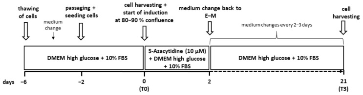
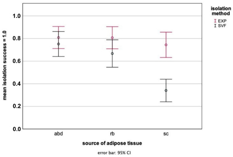
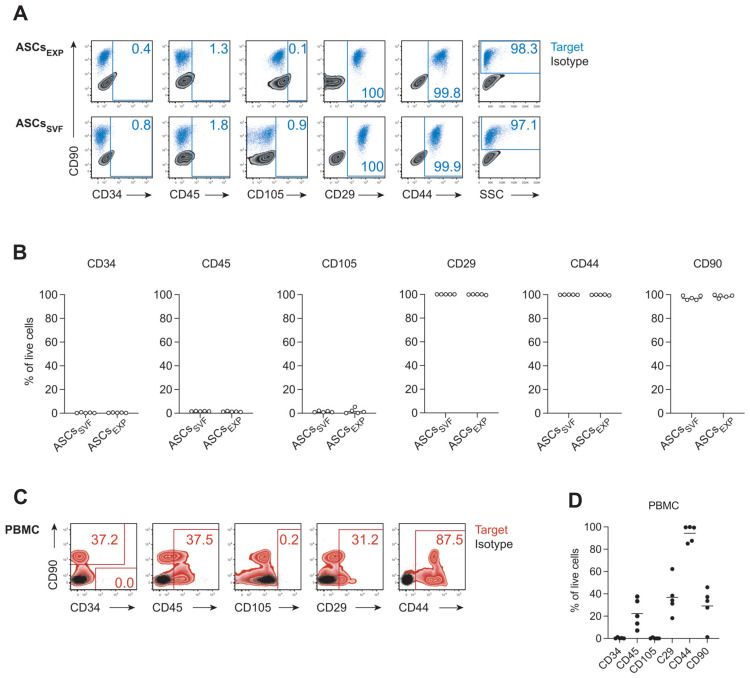
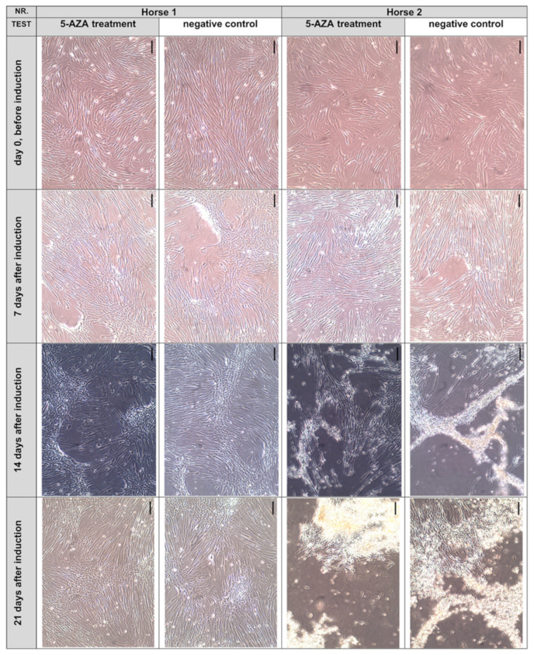
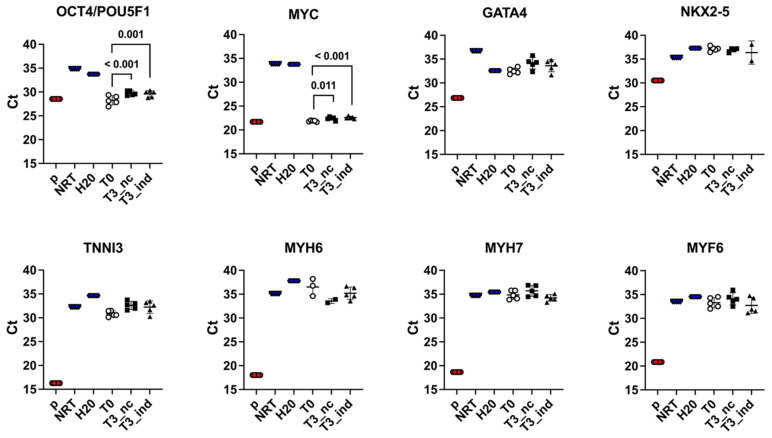
Physiological particularities of the equine heart justify the development of an in vitro model suitable for investigations of the species-specific equine cardiac electrophysiology. Adipose tissue-derived stromal/stem cells (ASCs) could be a promising starting point from which to develop such a cardiomyocyte (CM)-like cell model. Therefore, we compared abdominal, retrobulbar, and subcutaneous adipose tissue as sources for the isolation of ASCs applying two isolation methods: the collagenase digestion and direct explant culture. Abdominal adipose tissue was most suitable for the isolation of ASCs and both isolation methods resulted in comparable yields of CD45-/CD34-negative cells expressing the mesenchymal stem cell markers CD29, CD44, and CD90, as well as pluripotency markers, as determined by flow cytometry and real-time quantitative PCR. However, exposure of equine ASCs to 5-azacytidine (5-AZA), reportedly inducing CM differentiation from rats, rabbits, and human ASCs, was not successful in our study. More precisely, neither the early differentiation markers GATA4 and NKX2-5, nor the late CM differentiation markers TNNI3, MYH6, and MYH7 were upregulated in equine ASCs exposed to 10 µM 5-AZA for 48 h. Hence, further work focusing on the optimal conditions for CM differentiation of equine stem cells derived from adipose tissue, as well as possibly from other origins, are needed.
Keywords: adipose tissue differentiation; cardiomyocyte-like cells; horse; mesenchymal stem cells; preadipocytes.
Conflict of interest statement
The authors declare no conflict of interest. The funders had no role in the design of the study; in the collection, analyses, or interpretation of data; in the writing of the manuscript, or in the decision to publish the results.
Figures
Similar articles
-
Multilineage Differentiation Potential of Equine Adipose-Derived Stromal/Stem Cells from Different Sources.Animals (Basel). 2023 Apr 15;13(8):1352. doi: 10.3390/ani13081352. Animals (Basel). 2023. PMID: 37106915 Free PMC article.
-
[Differences between adipose-derived stem cells and mesenchymal stem cells in differentiation into cardiomyocytes].Sheng Li Xue Bao. 2008 Jun 25;60(3):341-7. Sheng Li Xue Bao. 2008. PMID: 18560724 Chinese.
-
Differentiation potential of human mesenchymal stem cells derived from adipose tissue and bone marrow to sinus node-like cells.Mol Med Rep. 2012 Jan;5(1):108-13. doi: 10.3892/mmr.2011.611. Epub 2011 Oct 3. Mol Med Rep. 2012. PMID: 21971826
-
Serum-free human MSC medium supports consistency in human but not in equine adipose-derived multipotent mesenchymal stromal cell culture.Cytometry A. 2018 Jan;93(1):60-72. doi: 10.1002/cyto.a.23240. Epub 2017 Sep 19. Cytometry A. 2018. PMID: 28926198
-
Adipose-derived mesenchymal stromal/stem cells: An update on their phenotype in vivo and in vitro.World J Stem Cells. 2014 Jul 26;6(3):256-65. doi: 10.4252/wjsc.v6.i3.256. World J Stem Cells. 2014. PMID: 25126376 Free PMC article. Review.
Cited by
-
Influence of the Anatomical Site on Adipose Tissue-Derived Stromal Cells' Biological Profile and Osteogenic Potential in Companion Animals.Vet Sci. 2023 Nov 24;10(12):673. doi: 10.3390/vetsci10120673. Vet Sci. 2023. PMID: 38133224 Free PMC article. Review.
-
Multilineage Differentiation Potential of Equine Adipose-Derived Stromal/Stem Cells from Different Sources.Animals (Basel). 2023 Apr 15;13(8):1352. doi: 10.3390/ani13081352. Animals (Basel). 2023. PMID: 37106915 Free PMC article.
References
-
- Rabkin S.W. Measurement of the QT interval: Lessons from thirty-two animal species for the correction of the QT interval by heart rate. Int. J. Clin. Cardiol. 2018;5:127. doi: 10.23937/2378-2951/1410127. - DOI
-
- Trachsel D.S., Tejada M.A., Groesfjeld Christensen V., Pedersen P.J., Kanters J.K., Buhl R., Calloe K., Klaerke D.A. Effects of trimethoprim-sulfadiazine and detomidine on the function of equine Kv 11.1 channels in a two-electrode voltage-clamp (TEVC) oocyte model. J. Vet. Pharmacol. Ther. 2018;41:536–545. doi: 10.1111/jvp.12502. - DOI - PubMed
-
- Pedersen P.J., Thomsen K.B., Flak J.B., Tejada M.A., Hauser F., Trachsel D., Buhl R., Kalbfleisch T., DePriest M.S., MacLeod J.N., et al. Molecular cloning and functional expression of the K+ channel KV7.1 and the regulatory subunit KCNE1 from equine myocardium. Res. Vet. Sci. 2017;113:79–86. doi: 10.1016/j.rvsc.2017.09.010. - DOI - PubMed
Grants and funding
LinkOut - more resources
Full Text Sources
Research Materials
Miscellaneous
