. 2022 Aug 19;12(16):2135.
doi: 10.3390/ani12162135.

MHCII Expression on Peripheral Blood Monocytes in Canine Lymphoma: An Impact of Glucocorticoids

Affiliations

MHCII Expression on Peripheral Blood Monocytes in Canine Lymphoma: An Impact of Glucocorticoids

Alicja Rzepecka et al. Animals (Basel). .

Abstract

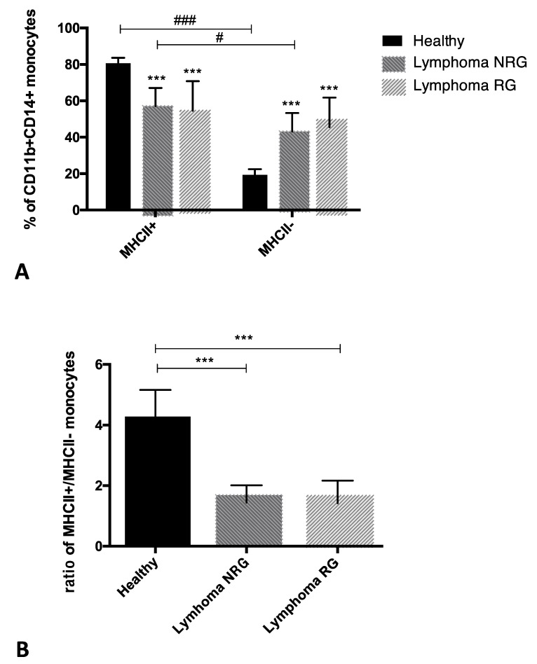
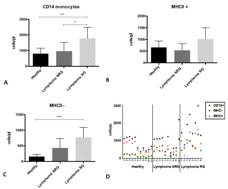
An increase in the percentage of monocytes with reduced HLA-DR expression and immunosuppressive properties has been reported in numerous human neoplastic diseases, including lymphoma. However, there are no analogous studies on phenotypical variations in the peripheral blood monocytes in dogs with lymphoma. The aim of this study was to determine the difference in the expression of the MHCII molecule on peripheral blood monocytes in dogs with lymphoma before any treatment (NRG) and in dogs that had previously received glucocorticoids (RG) in comparison to healthy dogs. Flow cytometry immunophenotyping of peripheral blood leukocytes was performed using canine-specific or cross-reactive antibodies against CD11b, CD14 and MHCII. In the blood of dogs with lymphoma (NRG and RG), compared to that of healthy ones, the MHCII+ and MHCII- monocytes ratio was changed due to an increase in the percentage of MHCII- monocytes. The number of MHCII- monocytes was significantly higher only in RG dogs compared to healthy ones, which might result from the release of these cells from the blood marginal pool due to the action of glucocorticoids. Our results encourage further studies to assess if changes in MHCII expression affect immune status in dogs with lymphoma.

Keywords: canine lymphoma; flow cytometry; glucocorticoids; immunosuppression; monocytes.


Conflict of interest statement

The authors declare no conflict of interest.

Figures

Figure 1
Figure 1
Representative dot plots depicting the gating strategy of canine peripheral blood leukocytes. (A) Forward scatter (FSC-hight) vs. side scatter (SSC-hight) dot plot showing leukocyte populations (population colors result from the regions set on the cytogram C); (B) debris and non-cellular elements were eliminated by setting a region defining CD45+ leukocytes (R1) on the SSC vs. CD45:APC dot plot; (C) leukocyte populations “(R1)” were analyzed on the CD45:APC/CD14:FITC dot plot in order to separate CD14+ monocytes (R2—red color) from lymphocytes (R3—blue color).
Figure 2
Figure 2
Representative dot plots depicting gating strategy of peripheral blood monocytes of dog with lymphoma. (A) Regions were set for all CD11b+ myeloid cells (R1) and for CD14+ monocytes (R2) on the CD14:APC vs. CD11b:PE double fluorescence dot plot; (B) distribution of all leukocytes (CD11b+ cells—red color) and (C) monocytes (from the R2 region—blue color) according to the expression of CD14 and MHCII. Bottom-row dot plots show isotype controls, separate for each Ab: anti-CD14 (A,B) and anti-MHCII (C). Controls allowed us to set the quadrants.
Figure 3
Figure 3
(A) The percentage of CD11b+CD14+ monocytes: MHCII+ and MHCII−, in the peripheral blood of healthy dogs (n = 11) and dogs with lymphoma: not receiving any drugs (NRG, n = 10) and receiving glucocorticoids (RG, n = 8). Arithmetic mean ± standard deviation. Significant differences between the percentage of MHCII+ and MHCII− monocytes among each group: # p < 0.05, ### p ≤ 0.001 (Wilcoxon) and for the percentage of MHCII+ or MHCII− monocytes in comparison to healthy dogs: *** p ≤ 0.001 (no significant differences between dogs with lymphoma NRG and RG) (Kruskal–Wallis and post hoc Dunn analysis). (B) The results are shown as the ratio of CD11b+CD14+ monocyte percentages: MHCII+ to MHCII−. Arithmetic mean ± standard deviation. Significant differences between groups: *** p ≤ 0.001 (Kruskal–Wallis and post hoc Dunn analysis).
Figure 4
Figure 4
The number of CD14+ monocytes in the peripheral blood of healthy dogs (n = 11) and dogs with lymphoma: not receiving any drugs (NRG, n = 10) and receiving glucocorticoids (RG, n = 8). (A) Total number of CD14+ monocytes per microliter of peripheral blood. The number of CD14+ cells was calculated based on their percentage in relation to all leukocytes (CD45+ cells) and white blood cell count. (B) The number of MHCII+ and (C) MHCII− monocyte subsets. Arithmetic mean ± standard deviation. Significant differences between groups: * p < 0.05, ** p ≤ 0.01, *** p ≤ 0.001 (Kruskal–Wallis and Dunn post hoc analysis). (D) The numbers of all evaluated cell populations in individual patients: MHCII− and MHCII+ monocytes as well as all CD14+ cell population.

References

    1. Holling T.M., Schooten E., van Den Elsen P.J. Function and regulation of MHC class II molecules in T-lymphocytes: Of mice and men. Hum. Immunol. 2004;65:282–290. doi: 10.1016/j.humimm.2004.01.005. - DOI - PubMed
    1. Ono S., Tsujimoto H., Matsumoto A., Ikuta S., Kinoshita M., Mochizuki H. Modulation of human leukocyte antigen-DR on monocytes and CD16 on granulocytes in patients with septic shock using hemoperfusion with polymyxin B-immobilized fiber. Am. J. Surg. 2004;188:150–156. doi: 10.1016/j.amjsurg.2003.12.067. - DOI - PubMed
    1. Yang H.M., Yu Y., Chai J.K., Hu S., Sheng Z.Y., Yao Y.M. Low HLA-DR expression on CD14+ monocytes of burn victims with sepsis, and the effect of carbachol in vitro. Burns. 2008;34:1158–1162. doi: 10.1016/j.burns.2008.01.026. - DOI - PubMed
    1. Winkler M.S., Rissiek A., Priefler M., Schwedhelm E., Robbe L., Bauer A., Zahrte C., Zoellner C., Kluge S., Nierhaus A. Human leucocyte antigen (HLA-DR) gene expression is reduced in sepsis and correlates with impaired TNFα response: A diagnostic tool for immunosuppression? PLoS ONE. 2017;12:e0182427. doi: 10.1371/journal.pone.0182427. - DOI - PMC - PubMed
    1. Weiss D.J., Welle M., Mortiz A., Walcheck B. Evaluation of leukocyte cell surface markers in dogs with septic and nonseptic inflammatory diseases. Am. J. Vet. Res. 2004;65:59–63. doi: 10.2460/ajvr.2004.65.59. - DOI - PubMed