. 2022 Jul 29;11(8):1137.
doi: 10.3390/biology11081137.

Myosin Transducer Inter-Strand Communication Is Critical for Normal ATPase Activity and Myofibril Structure

Affiliations

Myosin Transducer Inter-Strand Communication Is Critical for Normal ATPase Activity and Myofibril Structure

William A Kronert et al. Biology (Basel). .

Abstract

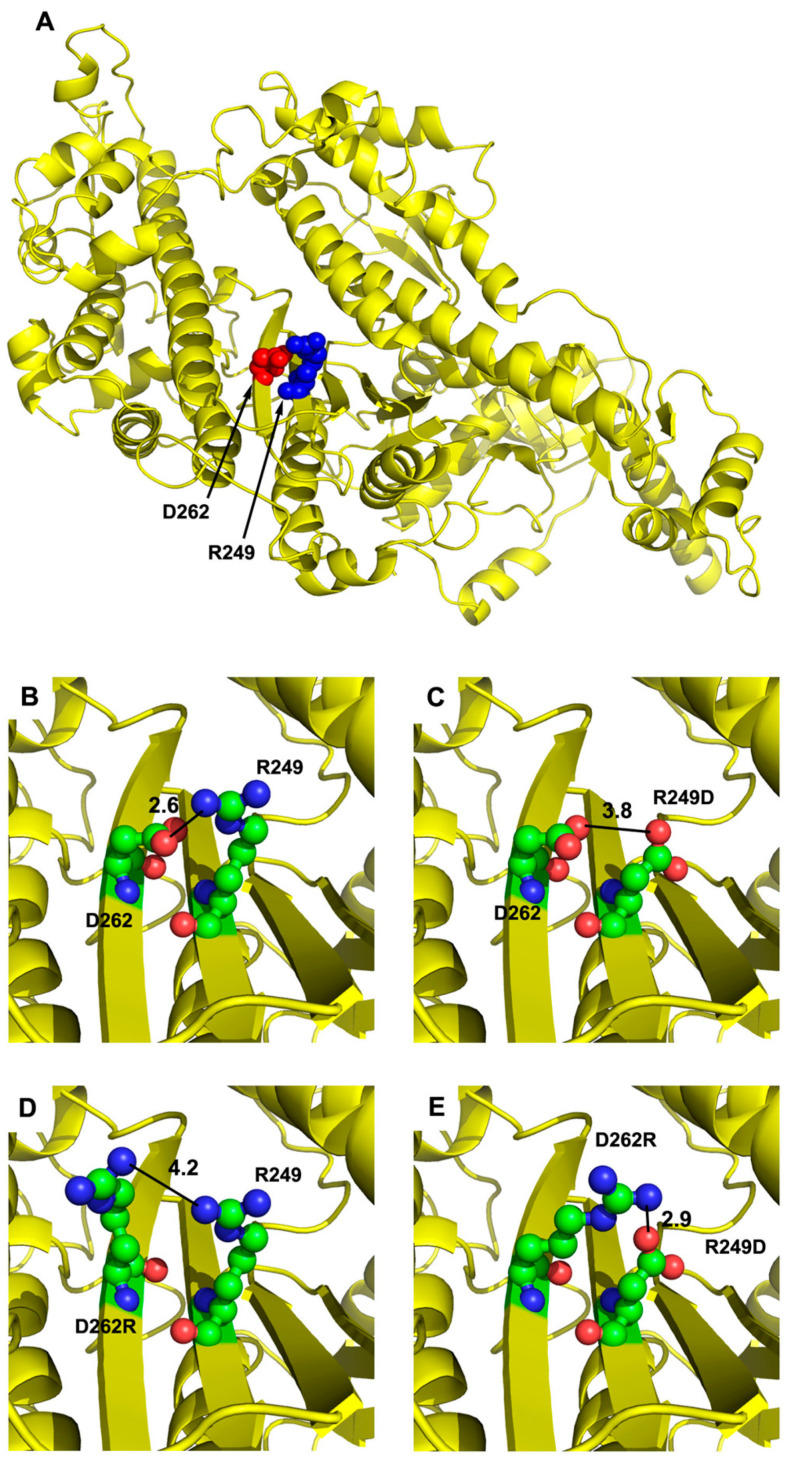
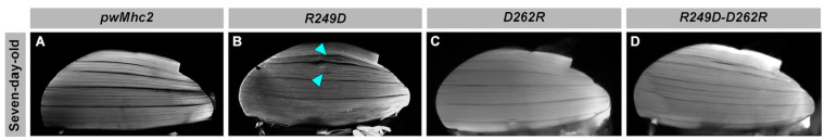
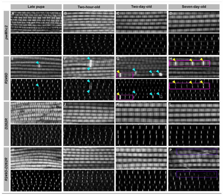
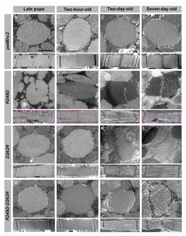
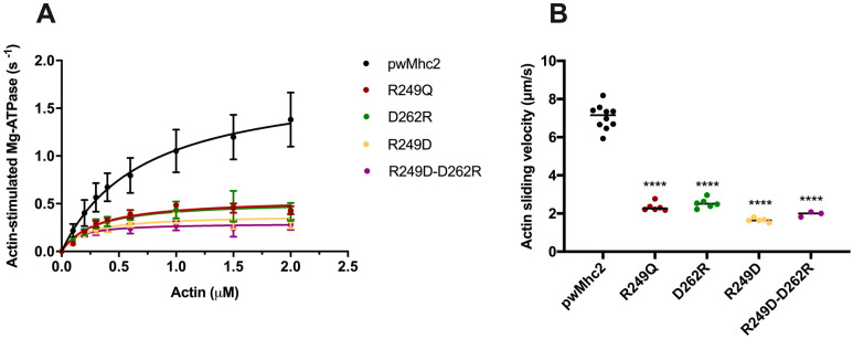
The R249Q mutation in human β-cardiac myosin results in hypertrophic cardiomyopathy. We previously showed that inserting this mutation into Drosophila melanogaster indirect flight muscle myosin yields mechanical and locomotory defects. Here, we use transgenic Drosophila mutants to demonstrate that residue R249 serves as a critical communication link within myosin that controls both ATPase activity and myofibril integrity. R249 is located on a β-strand of the central transducer of myosin, and our molecular modeling shows that it interacts via a salt bridge with D262 on the adjacent β-strand. We find that disrupting this interaction via R249Q, R249D or D262R mutations reduces basal and actin-activated ATPase activity, actin in vitro motility and flight muscle function. Further, the R249D mutation dramatically affects myofibril assembly, yielding abnormalities in sarcomere lengths, increased Z-line thickness and split myofibrils. These defects are exacerbated during aging. Re-establishing the β-strand interaction via a R249D/D262R double mutation restores both basal ATPase activity and myofibril assembly, indicating that these properties are dependent upon transducer inter-strand communication. Thus, the transducer plays an important role in myosin function and myofibril architecture.

Keywords: ATPase; Drosophila melanogaster; hypertrophic cardiomyopathy; myofibril; myosin; transducer.


Conflict of interest statement

The authors declare no conflict of interest.

Figures

Figure 1
Figure 1
Location and interaction of transducer residues R249 and D262 on the myosin molecule. (A) The human β-cardiac myosin motor domain (PDB: 4P7H) was used as a template for modeling the Drosophila indirect flight muscle myosin heavy chain sequence to determine the location of R249 (displayed as blue spheres) and D262 (red spheres). The R249 residue is located on the sixth strand and D262 is located on the seventh strand of the central β-sheet that is a portion of the myosin transducer. (B) The positively charged R249 residue interacts with negatively charged D262, with a 2.6 Å contact distance. (C) The R249D mutation disrupts the charged interaction with D262 due to their opposing charges, yielding an increase in contact distance to 3.8 Å. (D) The D262R mutation disrupts the charged interaction with R249 due to their opposing charges, yielding an increase in contact distance to 4.2 Å. (E) The R249D-D262R double mutation reestablishes the charged interaction with a contact distance of 2.9 Å.
Figure 2
Figure 2
Normal gross level structure of dorsolongitudinal IFM fibers of R249D, D262R and R249D-D262R mutant adults. Bisected thoraces were stained with phalloidin and imaged via fluorescence microscopy. Shown are seven-day-old adults with the following genotypes: (A) pwMhc2 wild-type control, (B) R249D, (C) D262R, (D) R249D-D262R double mutant. Fibers are of generally normal shape and size in all genotypes, indicating that there are no gross level defects, such as hypercontraction. However, R249D fibers show gaps (cyan arrowheads).
Figure 3
Figure 3
Confocal images of R249D, D262R and R249D-D262R myofibrils show defects in R249D that are rescued in the double mutant. Myofibrils from four developmental stages were imaged in longitudinal orientation to assess sarcomere structure. Muscles are stained for actin with fluorescent phalloidin and with an antibody to alpha-actinin, which localizes at Z-lines. pwMhc2 controls display regular Z- and M-lines within sarcomeric structures throughout development: (A) pwMhc2 late-stage pupa, (B) pwMhc2 two-hour-old adult, (C) pwMhc2 two-day-old adult, (D) pwMhc2 seven-day-old adult. (EH) Myofibrils from pwMhcR249D display minor assembly defects that are exacerbated during development. These include thick Z-line-like structures (cyan arrowheads), myofibril branching (magenta boxes), and occasionally both defects (yellow arrowheads). (IL) Myofibrils from pwMhcD262R show essentially wild-type sarcomere structures throughout development. (M) Myofibrils assemble normally in the pwMhcR249D-D262R double mutant at the late-pupal stage. (NO) Normal structure is maintained in young adults for pwMhcR249D-D262R. However, defects are present in seven-day-old pwMhcR249D-D262R myofibrils ((P), purple box). Thus, at this level of resolution, the D262R mutation rescues the various ultrastructural defects seen in pwMhcR249D myofibril assembly and early adult life. Scale bar, 5 µm.
Figure 4
Figure 4
Transmission electron microscopy of R249D, D262R and R249D-D262R myofibrils shows severe defects in R249D that are largely rescued in the double mutant. Transverse and longitudinal sections from (A) pwMhc2 late-stage pupae, (B) pwMhc2 two-hour-old adults, (C) pwMhc2 two-day-old adults, (D) pwMhc2 seven-day-old adults. Transverse sections display thick and thin filaments in a normal double-hexagonal pattern throughout development and aging. Longitudinal sections show regularly spaced Z- and M-lines. (E) Transverse and longitudinal sections from pwMhcR249D late-stage pupae display assembly defects with some disruption in thick and thin filament packing. Branching of the sarcomere is seen in longitudinal section (magenta box). (F) Transverse and longitudinal sections from pwMhcR249D two-hour-old adults show similar defects to late-stage pupae. Gaps in the sarcomere are seen in longitudinal section (magenta box). (G) Transverse and longitudinal sections of pwMhcR249D two-day-old adults display further disruption in muscle structure, with filament packing, myofibril branching and disrupted sarcomeres. (H) Transverse and longitudinal sections from pwMhcR249D seven-day-old adults display abnormal myofibril structure (magenta box), branching myofibrils and irregular Z-line placement that may correspond to thickened actin-containing structures (cyan arrowhead and adjacent Z-line structure to its right). (I,J) Transverse and longitudinal sections from pwMhcD262R late-stage pupae and two-hour-old adults resemble those of the transgenic control (Panels A and B), with normal hexagonal packing of thick and thin filaments and regular sarcomere structures. (K) Transverse section from pwMhcD262R two-day-old adult displays disruptions in thick and thin filament hexagonal packing. Sarcomere structure in longitudinal section remains relatively intact, with some myofilament gaps. (L) Transverse section from pwMhcD262R seven-day-old adult displays further disruptions in thick and thin filament hexagonal packing, with filaments loosely dispersed throughout the myofibril. Sarcomere structure remains relatively intact in longitudinal section, with some myofilament gaps and infiltration of Z-line material. (M,N) Transverse and longitudinal sections from pwMhcR249D-D262R late-stage pupae and two-hour-old adults resemble those of transgenic control (Panels A and B), with normal hexagonal packing of thick and thin filaments and normal sarcomere structures. This contrasts with abnormal myofibril morphology and filament packing in late-stage pupae and two-hour-old adults of pwMhcR249D (Panels E and F). (O) pwMhcR249D-D262R two-day-old adults display peripheral disruptions in thick and thin filament hexagonal packing in transverse section, with relatively normal sarcomere structure in longitudinal section. Myofibril morphology is less disrupted than in two-day-old pwMhcR249D adults (Panel G) or pwMhcD262R adults (Panel K). (P) Transverse and longitudinal sections from pwMhcR249D-D262R seven-day-old adults display more widespread disruption in thick and thin filament hexagonal packing. Sarcomere structure shows some disruption of M- and Z-lines. Myofibril morphology and filament packing in seven-day-old pwMhcR249D-D262R adults are less disrupted compared to pwMhcR249D (panel H) or pwMhcD262R seven-day-old adults (Panel L). Scale bar, 0.55 µm. M, M-line; Z, Z-line.
Figure 5
Figure 5
Actin-stimulated ATPase and actin sliding velocity values are reduced for R249Q, R249D, D262R and R249D-D262R myosin molecules. (A) Actin-stimulated Mg-ATPase levels are plotted relative to actin concentration for control (PwMhc2) and mutant myosins, after subtraction of basal Mg-ATPase values determined in the absence of actin. Each of the mutant myosins dramatically reduces Mg-ATPase activity relative to control (see Table 3 Vmax values) and decreases the concentration of actin needed to yield half-maximal ATPase activity (see Table 3 Km values). No statistically significant improvement is observed in myosin containing the R249D-D262R double mutation relative to the single mutations. (B) Actin sliding velocities calculated for control (PwMhc2) and mutant myosins. All mutations significantly decrease actin sliding velocities (****) and no improvement is observed in myosin containing the R249D-D262R double mutation relative to the single mutations (Table 3).

Similar articles

Cited by

References

    1. Sweeney H.L., Houdusse A. Structural and functional insights into the myosin motor mechanism. Annu. Rev. Biophys. 2010;39:539–557. doi: 10.1146/annurev.biophys.050708.133751. - DOI - PubMed
    1. Coureux P.D., Sweeney H.L., Houdusse A. Three myosin V structures delineate essential features of chemo-mechanical transduction. EMBO J. 2004;23:4527–4537. doi: 10.1038/sj.emboj.7600458. - DOI - PMC - PubMed
    1. Wulf S.F., Ropars V., Fujita-Becker S., Oster M., Hofhaus G., Trabuco L.G., Pylypenko O., Sweeney H.L., Houdusse A.M., Schroder R.R. Force-producing ADP state of myosin bound to actin. Proc. Natl. Acad. Sci. USA. 2016;113:E1844–E1852. doi: 10.1073/pnas.1516598113. - DOI - PMC - PubMed
    1. Pospich S., Sweeney H.L., Houdusse A., Raunser S. High-resolution structures of the actomyosin-V complex in three nucleotide states provide insights into the force generation mechanism. Elife. 2021;10:e73724. doi: 10.7554/eLife.73724. - DOI - PMC - PubMed
    1. Rosenzweig A., Watkins H., Hwang D.S., Miri M., McKenna W., Traill T.A., Seidman J.G., Seidman C.E. Preclinical diagnosis of familial hypertrophic cardiomyopathy by genetic analysis of blood lymphocytes. N. Engl. J. Med. 1991;325:1753–1760. doi: 10.1056/NEJM199112193252501. - DOI - PubMed

LinkOut - more resources