Review
. 2022 Aug 8;11(8):1191.
doi: 10.3390/biology11081191.

The Cell Autonomous and Non-Cell Autonomous Aspects of Neuronal Vulnerability and Resilience in Amyotrophic Lateral Sclerosis

Affiliations
Review

The Cell Autonomous and Non-Cell Autonomous Aspects of Neuronal Vulnerability and Resilience in Amyotrophic Lateral Sclerosis

Christoph Schweingruber et al. Biology (Basel). .

Abstract

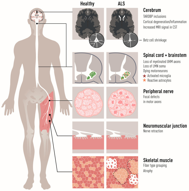
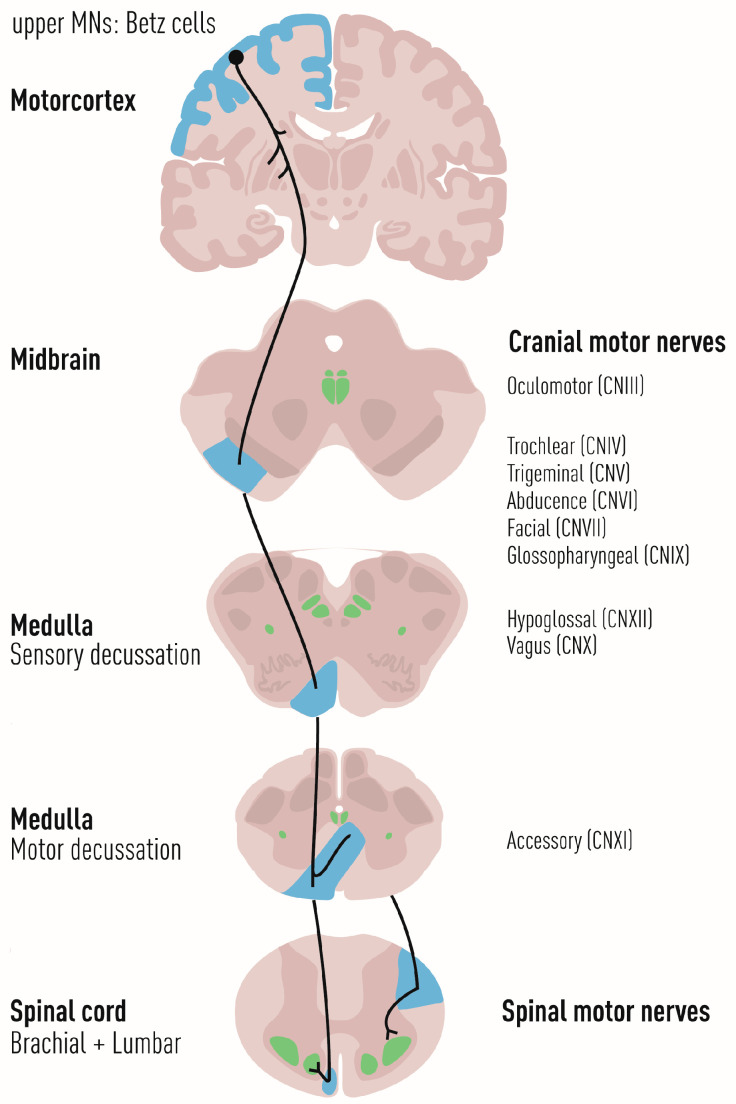
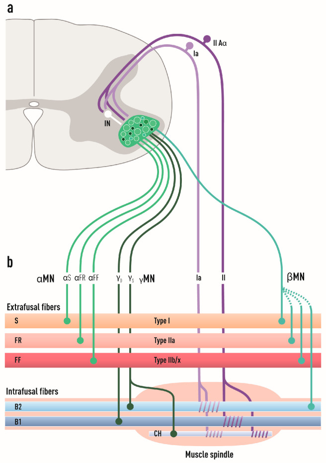
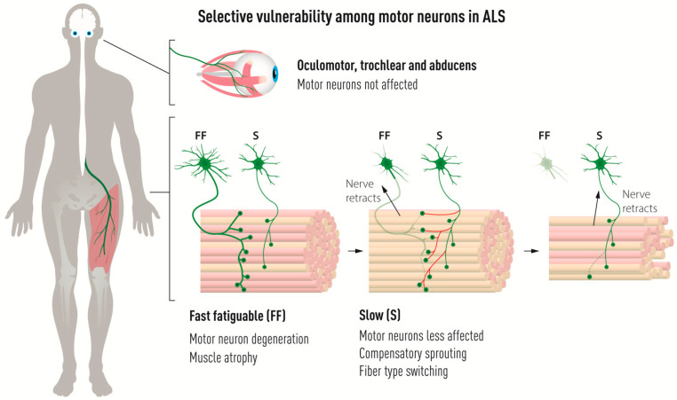
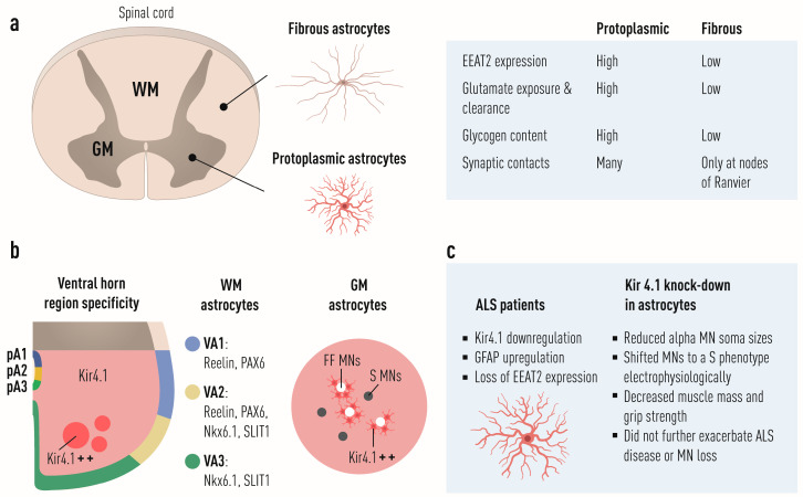
Amyotrophic lateral sclerosis (ALS) is defined by the loss of upper motor neurons (MNs) that project from the cerebral cortex to the brain stem and spinal cord and of lower MNs in the brain stem and spinal cord which innervate skeletal muscles, leading to spasticity, muscle atrophy, and paralysis. ALS involves several disease stages, and multiple cell types show dysfunction and play important roles during distinct phases of disease initiation and progression, subsequently leading to selective MN loss. Why MNs are particularly vulnerable in this lethal disease is still not entirely clear. Neither is it fully understood why certain MNs are more resilient to degeneration in ALS than others. Brain stem MNs of cranial nerves III, IV, and VI, which innervate our eye muscles, are highly resistant and persist until the end-stage of the disease, enabling paralyzed patients to communicate through ocular tracking devices. MNs of the Onuf's nucleus in the sacral spinal cord, that innervate sphincter muscles and control urogenital functions, are also spared throughout the disease. There is also a differential vulnerability among MNs that are intermingled throughout the spinal cord, that directly relate to their physiological properties. Here, fast-twitch fatigable (FF) MNs, which innervate type IIb muscle fibers, are affected early, before onset of clinical symptoms, while slow-twitch (S) MNs, that innervate type I muscle fibers, remain longer throughout the disease progression. The resilience of particular MN subpopulations has been attributed to intrinsic determinants and multiple studies have demonstrated their unique gene regulation and protein content in health and in response to disease. Identified factors within resilient MNs have been utilized to protect more vulnerable cells. Selective vulnerability may also, in part, be driven by non-cell autonomous processes and the unique surroundings and constantly changing environment close to particular MN groups. In this article, we review in detail the cell intrinsic properties of resilient and vulnerable MN groups, as well as multiple additional cell types involved in disease initiation and progression and explain how these may contribute to the selective MN resilience and vulnerability in ALS.

Keywords: amyotrophic lateral sclerosis; neurodegeneration; neurodegenerative cell metabolism; neuron.


Conflict of interest statement

The authors declare no conflict of interest. The funders had no role in reviewing the literature or in the writing of the manuscript.

Figures

Figure 1
Figure 1
The pathology of ALS encompasses all levels of the voluntary neuromuscular system and involves multiple cell types. The primary targets of ALS are motor neurons in the cortex, brainstem, and spinal cord. Neuropathological characteristics of most ALS cases are TDP43 inclusions in both neurons and glial cells. Neuroinflammation is raging throughout the nervous system with activated microglia and astrocytes seen wherever degeneration is occurring. The loss of upper and lower motor neurons is further evident from axonal degeneration and demyelination of the corticospinal tract as well as of the peripheral spinal nerves innervating skeletal muscles [2,3]. Denervation of neuromuscular junctions on affected skeletal muscle fibers have begun prior to clinical onset of any apparent motor deficit. While FF motor axons retract and degenerate, S motor neurons sprout and innervate endplates vacated by FF motor neurons. This new innervation results in fiber type switching and apparent grouping of muscle fiber types followed by atrophy of permanently denervated muscle fibers [4,5,6].
Figure 2
Figure 2
The upper motor system in humans and its connectivity with the lower motor system. Upper motor neurons are large pyramidal cells in the motor cortex, that are called Betz cells (blue). Their axons project through the corticospinal tract and innervate the nuclei of different lower motor neurons in the brainstem and spinal cord (green). Upper motor neurons are vulnerable to degeneration in ALS. Lower motor neurons are responsible for voluntary control of skeletal muscles and are vulnerable in ALS to different degrees.
Figure 3
Figure 3
The subdivision of lower somatic motor neurons and their connectivity with different muscle fiber types. (a) Lower motor neurons located in the ventral horn of the spinal cord innervate skeletal muscle fibers. They receive modulating signals from interneurons and also integrate inhibitory signals from afferent sensory neurons (Ia and II group IIAα) through the skeletal muscle that they both innervate. Although motor neuron somas are segregated according to muscle group in the motor column, the different motor neuron subtypes (alpha, beta, gamma) are intermingled. (b) Alpha motor neurons innervate extrafusal skeletal muscle fibers, which generate contractile forces. Alpha motor neurons are subdivided into fast-twitch fast fatigable (FF), fast-twitch fatigue-resistant (FR), and slow-twitch (S), which innervate type IIb/x, type IIa, and type I muscle fibers, respectively. Gamma motor neurons that are coactivated together with alpha motor neurons innervate intrafusal fibers that form stretch receptors in the form of muscle spindles and create a parallel pull of extra- and intrafusal fibers. The stretching of the spindle is detected by sensory neurons wrapping around the intrafusal fibers. Gamma motor neurons form two subtypes (dynamic and static) innervating intrafusal nuclear bag fibers (B1, B2) and nuclear chain fibers (CH). The dynamic units are required for quick adjustments to muscle tonus, whereas the static units are mainly involved in maintaining muscle tonus while posturing. Beta motor neurons innervate the muscle spindles and also have collaterals to the extrafusal fibers.
Figure 4
Figure 4
Selective vulnerability among somatic motor neurons in ALS. In ALS, somatic motor neurons of cranial motor neurons of the oculomotor, trochlear, and abducens nuclei, which regulate eye movement, remain unaffected until the end-stage of the disease. Among spinal motor neurons, there is a differential vulnerability with fast fatigable (FF) motor neurons being highly vulnerable and degenerating before symptoms onset, with resulting muscle atrophy. Slow (S) motor neurons are affected later in the disease and compensate for the loss of FF motor neurons by sprouting and innervating vacated endplates that were previously occupied by FF motor neurons. This results in fiber type switching of muscle from fast to slow. Eventually, S motor neurons are also affected and their NMJs with muscle are disrupted and their axons retract from muscle with resulting muscle weakness and atrophy.
Figure 5
Figure 5
Dorsoventral astrocyte diversity in the spinal cord and implications for differential spinal motor neuron vulnerability in ALS. (a) Astrocytes are subdivided into fibrous, in the white matter (WM), and protoplasmic, in the gray matter (GM). These differ in their EEAT2 expression, glutamate exposure and clearance, glycogen content, and synaptic contacts. (b) Astrocytes within the WM show regional specificity and arise from three progenitor domains, p1, p2, and p3. These subtypes can be identified on the basis of their expression of PAX6, NKX6.1, and the cell surface markers, reelin and SLIT1. Specifically, dorsally located VA1 astrocytes, derived from the p1 domain, express PAX6 and reelin; ventrally located VA3 astrocytes, derived from the p3 domain, express NKX6.1 and SLIT1,]; and intermediate WM-located VA2 astrocytes, derived from the p2 domain, express PAX6, NKX6.1, reelin, and SLIT1 [136]. GM astrocytes also show regional specificity with clear differences in KIR4.1 (KCNJ10) expression, with ventral horn astrocytes exhibiting higher expression of KIR4.1 than dorsal horn astrocytes. Within the ventral horn, WM astrocytes surrounding FF MNs show the highest level of KIR4.1 (KIR4.1++) and those surrounding S MNs show a lower level of KIR4.1. (c) ALS patient astrocytes show decreased expression of KIR4.1, GFAP upregulation and loss of EEAT2 expression. Removal of KIR4.1 from astrocytes in mice affected αMN soma sizes and shifted electrophysiological properties to a more slow-like MN phenotype with an accompanying decrease in muscle mass and grip strength. Overexpression of KIR4.1 specifically in astrocytes was sufficient to increase the size of both FF and S MNs. However, loss of KIR4.1 in astrocytes in the SOD1G93A mouse did not further exacerbate MN death.

References

    1. Swinnen B., Robberecht W. The phenotypic variability of amyotrophic lateral sclerosis. Nat. Rev. Neurol. 2014;10:661–670. doi: 10.1038/nrneurol.2014.184. - DOI - PubMed
    1. Riva N., Iannaccone S., Corbo M., Casellato C., Sferrazza B., Lazzerini A., Scarlato M., Cerri F., Previtali S.C., Nobile-Orazio E., et al. Motor nerve biopsy: Clinical usefulness and histopathological criteria. Ann. Neurol. 2011;69:197–201. doi: 10.1002/ana.22110. - DOI - PubMed
    1. Riva N., Clarelli F., Domi T., Cerri F., Gallia F., Trimarco A., Brambilla P., Lunetta C., Lazzerini A., Lauria G., et al. Unraveling gene expression profiles in peripheral motor nerve from amyotrophic lateral sclerosis patients: Insights into pathogenesis. Sci. Rep. 2016;6:39297. doi: 10.1038/srep39297. - DOI - PMC - PubMed
    1. Sharp P.S., Dick J.R.T., Greensmith L. The effect of peripheral nerve injury on disease progression in the SOD1 (G93A) mouse model of amyotrophic lateral sclerosis. Neuroscience. 2005;130:897–910. doi: 10.1016/j.neuroscience.2004.09.069. - DOI - PubMed
    1. Morris C.J. Human skeletal muscle fibre type grouping and collateral re-innervation. J. Neurol. Neurosurg. Psychiatry. 1969;32:440–444. doi: 10.1136/jnnp.32.5.440. - DOI - PMC - PubMed

LinkOut - more resources