Review
. 2022 Aug 22;11(8):1245.
doi: 10.3390/biology11081245.

New Insights into the Diversity of Branchiomeric Muscle Development: Genetic Programs and Differentiation

Affiliations
Review

New Insights into the Diversity of Branchiomeric Muscle Development: Genetic Programs and Differentiation

Imadeldin Yahya et al. Biology (Basel). .

Abstract

Branchiomeric skeletal muscles are a subset of head muscles originating from skeletal muscle progenitor cells in the mesodermal core of pharyngeal arches. These muscles are involved in facial expression, mastication, and function of the larynx and pharynx. Branchiomeric muscles have been the focus of many studies over the years due to their distinct developmental programs and common origin with the heart muscle. A prerequisite for investigating these muscles' properties and therapeutic potential is understanding their genetic program and differentiation. In contrast to our understanding of how branchiomeric muscles are formed, less is known about their differentiation. This review focuses on the differentiation of branchiomeric muscles in mouse embryos. Furthermore, the relationship between branchiomeric muscle progenitor and neural crest cells in the pharyngeal arches of chicken embryos is also discussed. Additionally, we summarize recent studies into the genetic networks that distinguish between first arch-derived muscles and other pharyngeal arch muscles.

Keywords: branchiomeric muscles; differentiation; mouse embryo; neural crest cells.


Conflict of interest statement

The authors declare that the research was conducted in the absence of any commercial or financial relationships that could be construed as a potential conflict of interest.

Figures
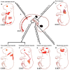
Figure 1
Figure 1
Summary of the embryonic origins of the branchiomeric and trunk muscles. (A) The branchiomeric muscle anlagen and second heart field progenitor cells originate from the cardiopharyngeal mesoderm that colonizes the core of the pharyngeal arches. (B) The somitic mesoderm gives rise to trunk and limb muscles. (C) The cardiopharyngeal mesoderm of arches 1–6 gives rise to cardiac muscle. (D) The cardiopharyngeal mesoderm of the first pharyngeal arch gives rise to mastication muscles. (E) The cardiopharyngeal mesoderm of the second pharyngeal arch gives rise to facial expression muscles. The caudal cardiopharyngeal mesoderm gives rise to the striated muscles of the esophagus (F) and non-somitic neck muscles (G). Retrieved from https://app.biorender.com/biorender-templates (accessed on 7 July 2022).
Figure 2
Figure 2
Summary of the distinct genetic program that governs myogenesis in the branchiomeric and trunk muscles. (A,B) Model of the genetic networks involved in branchiomeric and trunk muscles. The transcription factors Pitx2, Tbx1, Islet1, and MyoR set up the cardiopharyngeal mesoderm as a skeletal/heart-muscle-competent tissue. Pitx2 is required for the first pharyngeal arch muscle specification by modulating pre-myogenic markers (Tbx1, Capsulin, and MyoR). These genes are required for the activation of myogenic regulatory factors (MyoD and Myf5). The onset of Myf5 and MyoD commits branchiomeric muscle specification. MyoD directly activates genes implicated in keeping myoblasts in a proliferative state, whereas MyoG has antiproliferative activity through the activation of genes that block cell proliferation, promoting cell cycle exit and entry into terminal differentiation [45]. Pitx2 regulates the expression of Islet1, a second heart field marker. Tbx1 is required for the specification of second and caudal pharyngeal muscles. Tbx1 also regulates the expression of Islet1. Initiation of the myogenic program in the trunk and limb is regulated by Pax3, which is not expressed in the cardiopharyngeal mesoderm. BMP4 signals promote cardiogenesis in the head region and block skeletal muscle myogenesis in both the trunk and branchiomeric muscles. Wnt signaling inhibits branchiomeric muscle formation and initiates myogenesis in the trunk region. Antagonists of BMP4 (Noggin and Cremlin) and Wnt (Frzb) signals block cardiogenesis and induce the formation of branchiomeric muscle. PA, pharyngeal arch; ov, otic vesicle. Retrieved from https://app.biorender.com/biorender-templates (accessed on 7 July 2022).
Figure 3
Figure 3
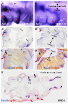
The emergence of the non-ectomesenchymal neural crest in the pharyngeal arches. (A,B) Analysis of Sox10 expression in developing chicken embryos using whole-mount in situ hybridization. Lateral views of the left side of chicken embryo stage HH20 and HH23. Sox10 was detected in the otic vesicle, facial ganglion, trigeminal ganglion, and first pharyngeal arch (blue arrow). Sox10 was first detected in the second pharyngeal arch at stage HH23 (facial nerve, black arrow). (C,D) Analysis of Myf5 expression using whole-mount in situ hybridization. (E) Frontal and sagittal (F) sections of a chicken embryo at the level of the second pharyngeal arch were hybridized with the MyoD probe, followed by immunostaining using an HNK1 antibody. Myf5 (white arrows) and MyoD (black arrow) mark the mesodermal core of the pharyngeal arches. In (E,F), cranial nerves in the first and second pharyngeal arches are revealed by HNK1 staining (red arrows). PA1, first pharyngeal arch; PA2, second pharyngeal arch; ov, otic vesicle; V, trigeminal ganglion; VII, facial ganglion.
Figure 4
Figure 4
Ap2α marks ectomesenchymal neural crest cells in the first and second pharyngeal arches. (A,B) Analysis of Ap2α expression using whole-mount in situ hybridization. Lateral views of stages HH21-22 and HH23-24. (A’,B’) Adjacent vibratome frontal sections of the chicken embryos in (A,B) at the level of first and second pharyngeal arches. (C,D) Immunostaining for Desmin on the same frontal sections as in (A’,B’) after whole-mount in situ hybridization. The mesodermal core was visualized with Desmin antibody (black arrows). (E) The frontal section shows double whole-mount in situ hybridization for MyoD (blue) and Ap2α (red). Note that Ap2α marks the ectomesnchymal neural crest (red arrows) and MyoD marks the mesodermal core of the pharyngeal arches (black arrows). PA1, first pharyngeal arch; PA2, second pharyngeal arch; ov, otic vesicle.
Figure 5
Figure 5
The emergence of myosin heavy chain during the development of the mouse branchiomeric muscle. (AD) Analysis of myosin heavy chain (MyHC) expression using whole-mount in situ hybridization. Lateral views of E10.5, E11.5, E12.5, and E13.5 mouse embryos. The MyHC transcripts emerged in the first arch-derived muscle anlagen (white arrow in (A)) before the second and caudal brachial-derived muscle anlagen. Note: a-trap, acromiotrapezius; au, auricularis; bu; buccinator; fr, frontalis; ma, masseter; PA1, first pharyngeal arch; PA2, second pharyngeal arch; qu, quadratus labii; snm, somitic neck muscle; s-trap, spinotrapezius; te, temporalis, zy, zygomaticus.

Similar articles

Cited by

References

    1. Lescroart F., Kelly R.G., Le Garrec J.F., Nicolas J.F., Meilhac S.M., Buckingham M. Clonal analysis reveals common lineage relationships between head muscles and second heart field derivatives in the mouse embryo. Development. 2010;137:3269–3279. doi: 10.1242/dev.050674. - DOI - PubMed
    1. Nogueira J.M., Hawrot K., Sharpe C., Noble A., Wood W.M., Jorge E.C., Goldhamer D.J., Kardon G., Dietrich S. The emergence of Pax7-expressing muscle stem cells during vertebrate head muscle development. Front. Aging Neurosci. 2015;7:62. doi: 10.3389/fnagi.2015.00062. - DOI - PMC - PubMed
    1. Buckingham M., Vincent S.D. Distinct and dynamic myogenic populations in the vertebrate embryo. Curr. Opin. Genet. Dev. 2009;19:444–453. doi: 10.1016/j.gde.2009.08.001. - DOI - PubMed
    1. Sambasivan R., Kuratani S., Tajbakhsh S. An eye on the head: The development and evolution of craniofacial muscles. Development. 2011;138:2401–2415. doi: 10.1242/dev.040972. - DOI - PubMed
    1. Buckingham M., Rigby P.W.J. Gene Regulatory Networks and Transcriptional Mechanisms that Control Myogenesis. Dev. Cell. 2014;28:225–238. doi: 10.1016/j.devcel.2013.12.020. - DOI - PubMed

LinkOut - more resources