Review
. 2022 Aug 15;11(8):1105.
doi: 10.3390/antibiotics11081105.

Chelation in Antibacterial Drugs: From Nitroxoline to Cefiderocol and Beyond

Affiliations
Review

Chelation in Antibacterial Drugs: From Nitroxoline to Cefiderocol and Beyond

Davorka Repac Antić et al. Antibiotics (Basel). .

Abstract

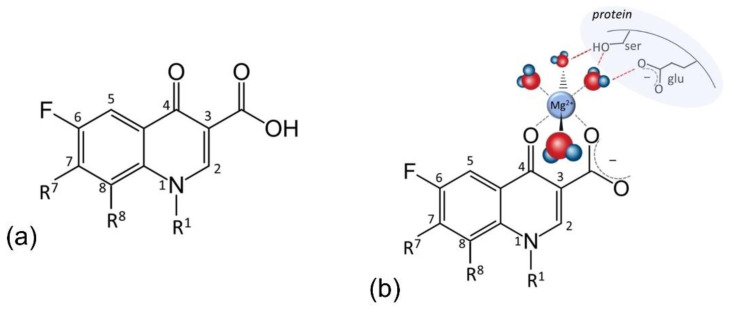
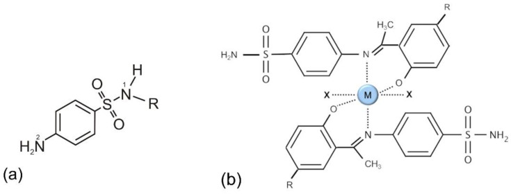
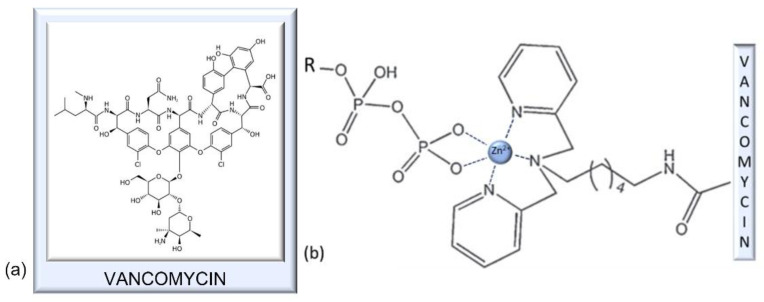
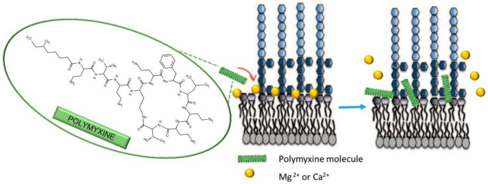
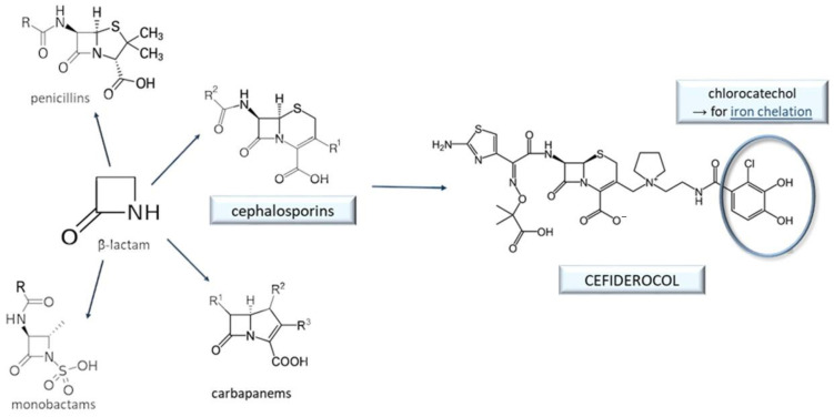
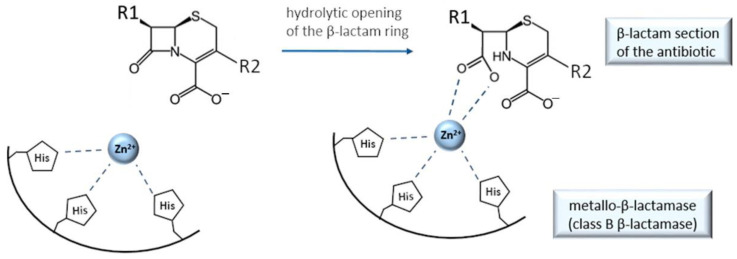
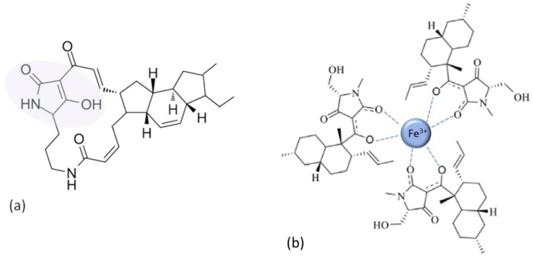
In the era of escalating antimicrobial resistance, the need for antibacterial drugs with novel or improved modes of action (MOAs) is a health concern of utmost importance. Adding or improving the chelating abilities of existing drugs or finding new, nature-inspired chelating agents seems to be one of the major ways to ensure progress. This review article provides insight into the modes of action of antibacterial agents, class by class, through the perspective of chelation. We covered a wide scope of antibacterials, from a century-old quintessential chelating agent nitroxoline, currently unearthed due to its newly discovered anticancer and antibiofilm activities, over the commonly used antibacterial classes, to new cephalosporin cefiderocol and a potential future class of tetramates. We show the impressive spectrum of roles that chelation plays in antibacterial MOAs. This, by itself, demonstrates the importance of understanding the fundamental chemistry behind such complex processes.

Keywords: antibacterial drugs; antibacterial modes of action (MOAs); chelation; nitroxoline.


Conflict of interest statement

The authors declare no conflict of interest.

Figures

Figure 1
Figure 1
(a) Structure of nitroxoline (5-nitro-8-hydroxyquinoline, NIT); (b) example of NIT−Cu2+ chelate, showing two possible chelating sites.
Figure 2
Figure 2
SEM images of E. faecalis on urinary catheter surface (a) before and (b) after nitroxoline treatment, showing the areas of biofilm destruction. Samples were coated with Pd before analysis on a Hitachi S–3600N Scanning Electron Microscope. Image taken at 13.3 k magnification (scale bar represents 10 μm).
Figure 3
Figure 3
(a) Structure of tetracycline. The shaded area emphasizes the part relevant for chelating ability. (b) An example of a simple tetracycline–Mg chelate.
Figure 4
Figure 4
Binding of the TC molecule (tigecycline) to phosphate group (P) oxygens of the rRNA backbone in the bacterial 30S ribosomal subunit. The binding is done both directly and via Mg2+ ion(s). The image is inspired by ref. [93].
Figure 5
Figure 5
(a) General structure of fluoroquinolones. (b) Chelation of Mg2+ by a fluoroquinolone molecule (moxifloxacin), as the core of their mode of action. Two water molecules serve as a bridge to bacterium topoisomerase IV (hydrogen bonds indicated by red dashed line), blocking phosphotyrosine from approaching the other Mg2+ at the enzyme’s active site. Hydrogen bonds with DNA strand also present are not shown for simplicity. Inspired by ref. [45].
Figure 6
Figure 6
(a) General structure of sulfonamides. (b) Chelation is not relevant in the classical sulfonamide mode of action, but the chelates of new-generation sulfonamide molecules with metal (M) cations (M = Co2+, Ni2+, Cu2+, Zn2+, and VO2+) demonstrate antibacterial and antioxidative activities; inspired by ref. [49].
Figure 7
Figure 7
(a) Structure of vancomycin, a glycopeptide antibiotic. (b) Zinc chelate of dipicolyl–vancomycin conjugate and pyrophosphate groups of cell-wall lipids. Inspired by ref. [50].
Figure 8
Figure 8
One of the proposed modes of action of polymyxins on Gram-negative bacteria. Due to higher affinity to membrane lipopolysaccharides (LPS) compared to native Mg2+ and Ca2+, polymyxine molecules displace the cations from LPS-cation chelates (crucial for membrane stability), thus disrupting the physical integrity of the membrane. Inspired by ref. [55].
Figure 9
Figure 9
Structure of bacitracin.
Figure 10
Figure 10
Generic structures of (a) macrolides and (b) lincosamides.
Figure 11
Figure 11
Classification of the β-lactam antibacterials. Cefiderocol, a member of the cephalosporin subgroup, contains a catechol moiety that enables it to chelate Fe3+ ions and thus use the bacterial ferric ion transport system to enter the bacterial cell (the Trojan horse approach).
Figure 12
Figure 12
Simplified scheme of metallo-β-lactamase catalytic mechanism, depicting the role of chelation in disabling the action of β-lactam antibiotics (inspired by ref. [71]). For clarity, only one out of two Zn2+ ions are shown.
Figure 13
Figure 13
(a) Ikarugamycin, a member of the tetramate class and the polycyclic tetramate macrolactams (PTM) subgroup. Tetramate moiety is emphasized in the shaded area. (b) An example of the tetramate chelate, comprising three tetramate (equisetin) molecules as bidentate ligands chelating Fe3+ cation. Inspired by ref. [72].

References

    1. Hutchings M.I., Truman A.W., Wilkinson B. Antibiotics: Past, Present and Future. Curr. Opin. Microbiol. 2019;51:72–80. doi: 10.1016/j.mib.2019.10.008. - DOI - PubMed
    1. Barzic A.I., Ioan S. Antibacterial Drugs—From Basic Concepts to Complex Therapeutic Mechanisms of Polymer Systems. In: Bobbarala V., editor. Concepts, Compounds and the Alternatives of Antibacterials. IntechOpen; London, UK: 2015.
    1. Kapoor G., Saigal S., Elongavan A. Action and Resistance Mechanisms of Antibiotics: A Guide for Clinicians. J. Anaesthesiol. Clin. Pharmacol. 2017;33:300. doi: 10.4103/joacp.JOACP_349_15. - DOI - PMC - PubMed
    1. Blanusa M., Varnai V.M., Piasek M., Kostial K. Chelators as Antidotes of Metal Toxicity: Therapeutic and Experimental Aspects. Curr. Med. Chem. 2005;12:2771–2794. doi: 10.2174/092986705774462987. - DOI - PubMed
    1. Palmer L.D., Skaar E.P. Transition Metals and Virulence in Bacteria. Annu. Rev. Genet. 2016;50:67–91. doi: 10.1146/annurev-genet-120215-035146. - DOI - PMC - PubMed

LinkOut - more resources