Review
. 2022 Aug 11;9(8):1210.
doi: 10.3390/children9081210.

From Fetal to Neonatal Neuroimaging in TORCH Infections: A Pictorial Review

Affiliations
Review

From Fetal to Neonatal Neuroimaging in TORCH Infections: A Pictorial Review

Giulia Lucignani et al. Children (Basel). .

Abstract

Congenital infections represent a challenging and varied clinical scenario in which the brain is frequently involved. Therefore, fetal and neonatal neuro-imaging plays a pivotal role in reaching an accurate diagnosis and in predicting the clinical outcome. Congenital brain infections are characterized by various clinical manifestations, ranging from nearly asymptomatic diseases to syndromic disorders, often associated with severe neurological symptoms. Brain damage results from the complex interaction among the infectious agent, its specific cellular tropism, and the stage of development of the central nervous system at the time of the maternal infection. Therefore, neuroradiological findings vary widely and are the result of complex events. An early detection is essential to establishing a proper diagnosis and prognosis, and to guarantee an optimal and prompt therapeutic perinatal management. Recently, emerging infective agents (i.e., Zika virus and SARS-CoV2) have been related to possible pre- and perinatal brain damage, thus expanding the spectrum of congenital brain infections. The purpose of this pictorial review is to provide an overview of the current knowledge on fetal and neonatal brain neuroimaging patterns in congenital brain infections used in clinical practice.

Keywords: CMV; MRI; SARS-CoV-2; TORCH; congenital infection; fetal imaging; neonatal imaging.


Conflict of interest statement

The authors of this manuscript declare that they have no competing interests.

Figures

Figure 1
Figure 1
Cerebral US performed in neonate affected by CMV, showing periventricular cysts (dotted white arrows in (A,B)) and subependimal hemorrhage (black asterisks in (C)) evolving in periventricular cysts (white asterisks in (D). Cerebral US performed in neonate affected by CMV, showing frontal calcifications in the coronal plane (white circles in (E)) and calcifications of the lenticulo-striatal arterioles in the sagittal plane (black arrow in (F)).
Figure 2
Figure 2
Fetus of 25 weeks of gestational age showing diffuse ventricular ectasis (E,F), cysternal and peri-encephalic spaces (BF) coexisting with periventricular cysts (black arrow in (A)), and polymicrogyria (B,C). Courtesy of Mónica Rebollo Polo, Hospital Sant Joan de Déu, Barcelona, Spain.
Figure 3
Figure 3
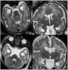
Fetuses of: 23 weeks of gestation age, showing polymicrogyria (A,B); 25 weeks of gestation age, showing cerebellar hypoplasia and polymicrogyria (C,D); 32 weeks of gestation age, showing white matter alterations (E,F).
Figure 4
Figure 4
US (A) and MRI (B) performed in one-month-old neonate affected by CMV showing a coarse frontal calcification in the anterior coronal plane and on axial GE T2* sequence, respectively. Six-month-old child showing multiple and bilateral periventricular calcifications on axial CT (C).
Figure 5
Figure 5
MRI performed in neonate affected by CMV in the first gestational trimester showing calcifications and cysts on axial (AC) and sagittal (D,H) T2WI and on axial T1WI (EG).
Figure 6
Figure 6
MRI performed in neonate affected by CMV in the first gestational trimester presenting with purpura, hepatosplenomegaly, coagulation, and neurological deficits, showing cerebellar and occipital hemorrhage coexisting with a subdural hematoma on coronal (A,B) and sagittal (C,D) T1WI and hemoventricle on coronal (E,F) and axial (G) T2WI and on axial T1WI (H).
Figure 7
Figure 7
Seventeen-month-old child affected by CMV and presenting with post-natal deafness and neurocognitive retardation. MRI showed mesial temporal cysts on axial FLAIR (A,B), coronal T2WI images (C,F) and bilateral periventricular white matter alterations on axial FLAIR images (D,E).
Figure 8
Figure 8
Four-day-old neonate affected by Toxoplasmosis and undergoing the cerebral US in the coronal plane, showing massive supratentorial ventricular dilatation (AC) and linear subependymal calcifications (A,B).
Figure 9
Figure 9
Pediatric patient affected by congenital CMV, undergoing MRI at four days of birth (AC) and CT at six months of life (DF), showing massive supratentorial ventricular dilatation and linear subependimal calcifications.
Figure 10
Figure 10
Fetus at 29 weeks gestational age affected by Toxoplasma, showing destruent frontal and insular white matter lesions (black arrow in (A,B)). Fetus of 34 weeks of gestation age affected by Toxoplasma, showing ventricular dilatation coexisting with destruent frontal white matter lesions (black arrow in (C,D)).
Figure 11
Figure 11
Twenty-day-old neonate affected by Aicardi-Goutieres type 1 syndromes, namely a pseudo-TORCH disease, showing white matter alterations, calcifications, and cystic vacuolization on US (A,D), CT (B,E), and MRI (C,F).
Figure 12
Figure 12
Fetal MRI performed on a fetus affected by Rubella at 27 weeks of gestational age, showing bilateral temporal cysts (black arrows in (A,B)), and subependymal cysts (white arrows in (B)). Neonatal MRI performed on the same patient at six days after birth, showing temporal cysts (black arrows in (C,D)) ventricular dilatation (black asterisks in (C,D)), and subependymal cysts (white arrow in (D)).
Figure 13
Figure 13
MRI of neonate affected by herpes virus type 1 (AD) presenting with seizures and right hemiparesis, showing cortical-subcortical signal alteration on pre-contrast T1WI (A), contrast enhancement on post-contrast T1WI and diffusion restriction on DWI/ADC (C,D) in the right parietal-insular region. Neonate affected by herpes virus type 2 undergoing US, showing cortical-subcortical hyperechogenicity in the right posterior temporal and left parietal regions (E), and undergoing MRI, showing diffusion restriction on DWI/ADC (F,G) and contrast-enhancement in the abovementioned areas (H).
Figure 14
Figure 14
Term neonate affected by SARS-CoV2 undergoing MRI at five days after birth, showing supratentorial and infratentorial hemoventricle coexisting with subarachnoid hemorrhage on coronal T2WI-T1WI (A,B) and sagittal T1 (C). Term neonate affected by SARS-CoV-2 undergoing MRI at three days after birth, showing bilateral frontal hemorrhagic foci and linear subependymal hemorrhage on axial T1WI (D) and DWI/ADC (E,F).
Figure 15
Figure 15
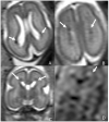
Fetus of 23 weeks of gestation age affected by parvovirus and presenting with anemia, cardiac failure, hydrops, and venous congestion. Fetal MRI shows hemorrhagic venous infarctions in deep medullary veins (white arrows in (AD)).

References

    1. Baldwin S., Whitley R. Teratogen update: Intrauterine herpes simplex virus infection. Teratology. 1989;39:1–10. doi: 10.1002/tera.1420390102. - DOI - PubMed
    1. Megli C.J., Coyne C.B. Infections at the maternal-fetal interface: An overview of pathogenesis and defense. Nat. Rev. Microbiol. 2022;20:67–82. doi: 10.1038/s41579-021-00610-y. - DOI - PMC - PubMed
    1. Nickerson J.P., Richner B., Santy K., Lequin M.H., Poretti A., Filippi C.G., Huisman T.A. Neuroimaging of pediatric intracranial infection—Part 2: TORCH, viral, fungal, and parasitic infections. J. Neuroimaging. 2012;22:e52–e63. doi: 10.1111/j.1552-6569.2011.00699.x. - DOI - PubMed
    1. Stegmann B.J., Carey J.C. TORCH Infections. Toxoplasmosis, Other (syphilis, varicella-zoster, parvovirus B19), Rubella, Cytomegalovirus (CMV), and Herpes infections. Curr. Womens Health Rep. 2002;2:253–258. - PubMed
    1. Colonna A.T., Buonsenso D., Pata D., Salerno G., Chieffo D.P.R., Romeo D.M., Faccia V., Conti G., Molle F., Baldascino A., et al. Long-Term Clinical, Audiological, Visual, Neurocognitive and Behavioral Outcome in Children with Symptomatic and Asymptomatic Congenital Cytomegalovirus Infection Treated with Valganciclovir. Front. Med. 2020;7:268. doi: 10.3389/fmed.2020.00268. - DOI - PMC - PubMed

LinkOut - more resources