

Convolutional Neural Network Techniques for Brain Tumor Classification (from 2015 to 2022): Review, Challenges, and Future Perspectives
- PMID: 36010200
- PMCID: PMC9406354
- DOI: 10.3390/diagnostics12081850
Convolutional Neural Network Techniques for Brain Tumor Classification (from 2015 to 2022): Review, Challenges, and Future Perspectives
Abstract
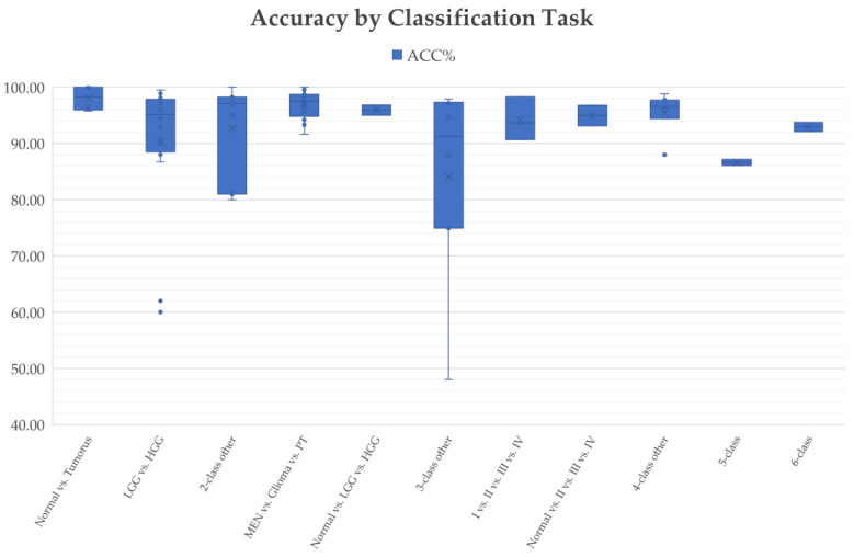
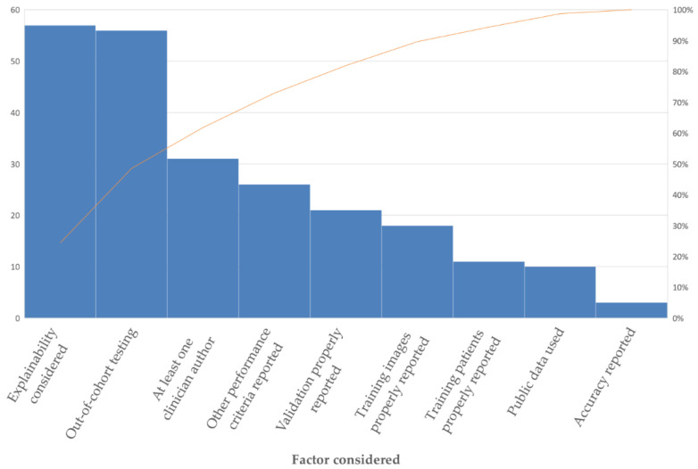
Convolutional neural networks (CNNs) constitute a widely used deep learning approach that has frequently been applied to the problem of brain tumor diagnosis. Such techniques still face some critical challenges in moving towards clinic application. The main objective of this work is to present a comprehensive review of studies using CNN architectures to classify brain tumors using MR images with the aim of identifying useful strategies for and possible impediments in the development of this technology. Relevant articles were identified using a predefined, systematic procedure. For each article, data were extracted regarding training data, target problems, the network architecture, validation methods, and the reported quantitative performance criteria. The clinical relevance of the studies was then evaluated to identify limitations by considering the merits of convolutional neural networks and the remaining challenges that need to be solved to promote the clinical application and development of CNN algorithms. Finally, possible directions for future research are discussed for researchers in the biomedical and machine learning communities. A total of 83 studies were identified and reviewed. They differed in terms of the precise classification problem targeted and the strategies used to construct and train the chosen CNN. Consequently, the reported performance varied widely, with accuracies of 91.63-100% in differentiating meningiomas, gliomas, and pituitary tumors (26 articles) and of 60.0-99.46% in distinguishing low-grade from high-grade gliomas (13 articles). The review provides a survey of the state of the art in CNN-based deep learning methods for brain tumor classification. Many networks demonstrated good performance, and it is not evident that any specific methodological choice greatly outperforms the alternatives, especially given the inconsistencies in the reporting of validation methods, performance metrics, and training data encountered. Few studies have focused on clinical usability.
Keywords: brain tumor classification; clinical application; clinical effectiveness; computer-aided diagnosis; convolutional neural network; deep learning; magnetic resonance imaging.
Conflict of interest statement
The authors declare no conflict of interest.
Figures

References
-
- Cancer Research UK [(accessed on 10 February 2022)]. Available online: https://www.cancerresearchuk.org/health-professional/cancer-statistics/s....
-
- Islami F., Ward E.M., Sung H., Cronin K.A., Tangka F.K.L., Sherman R.L., Zhao J.X., Anderson R.N., Henley S.J., Yabroff K.R., et al. Annual report to the nation on the status of cancer, part 1: National cancer statistics. JNCI J. Natl. Cancer Inst. 2021;113:1648–1669. doi: 10.1093/jnci/djab131. - DOI - PMC - PubMed
Publication types
Grants and funding
LinkOut - more resources
Full Text Sources
