

A Comprehensive Review of Machine Learning Used to Combat COVID-19
- PMID: 36010204
- PMCID: PMC9406981
- DOI: 10.3390/diagnostics12081853
A Comprehensive Review of Machine Learning Used to Combat COVID-19
Abstract
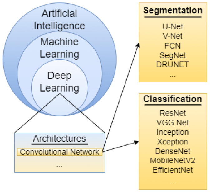
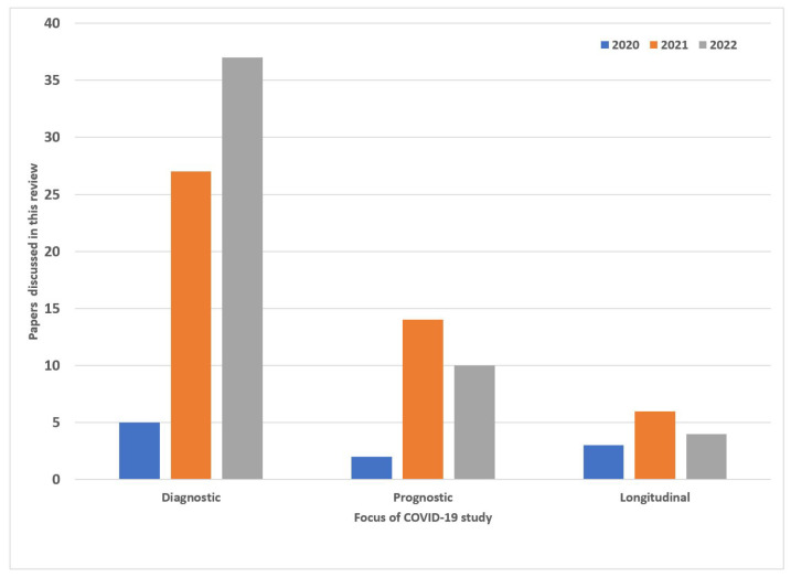
Coronavirus disease (COVID-19) has had a significant impact on global health since the start of the pandemic in 2019. As of June 2022, over 539 million cases have been confirmed worldwide with over 6.3 million deaths as a result. Artificial Intelligence (AI) solutions such as machine learning and deep learning have played a major part in this pandemic for the diagnosis and treatment of COVID-19. In this research, we review these modern tools deployed to solve a variety of complex problems. We explore research that focused on analyzing medical images using AI models for identification, classification, and tissue segmentation of the disease. We also explore prognostic models that were developed to predict health outcomes and optimize the allocation of scarce medical resources. Longitudinal studies were conducted to better understand COVID-19 and its effects on patients over a period of time. This comprehensive review of the different AI methods and modeling efforts will shed light on the role that AI has played and what path it intends to take in the fight against COVID-19.
Keywords: COVID-19 prognosis; CT scan; X-rays; deep learning; machine learning.
Conflict of interest statement
The authors declare no conflict of interest.
Figures

Similar articles
-
Artificial intelligence (AI) for medical imaging to combat coronavirus disease (COVID-19): a detailed review with direction for future research.Artif Intell Rev. 2022;55(2):1409-1439. doi: 10.1007/s10462-021-09985-z. Epub 2021 Apr 15. Artif Intell Rev. 2022. PMID: 33875900 Free PMC article.
-
Role of Machine Learning Techniques to Tackle the COVID-19 Crisis: Systematic Review.JMIR Med Inform. 2021 Jan 11;9(1):e23811. doi: 10.2196/23811. JMIR Med Inform. 2021. PMID: 33326405 Free PMC article. Review.
-
Combating COVID-19 Crisis using Artificial Intelligence (AI) Based Approach: Systematic Review.Curr Top Med Chem. 2024;24(8):737-753. doi: 10.2174/0115680266282179240124072121. Curr Top Med Chem. 2024. PMID: 38318824
-
A Systematic Review on the Use of AI and ML for Fighting the COVID-19 Pandemic.IEEE Trans Artif Intell. 2021 Mar 1;1(3):258-270. doi: 10.1109/TAI.2021.3062771. eCollection 2020 Dec. IEEE Trans Artif Intell. 2021. PMID: 35784006 Free PMC article.
-
Diagnosis of COVID-19 Using Machine Learning and Deep Learning: A Review.Curr Med Imaging. 2021;17(12):1403-1418. doi: 10.2174/1573405617666210713113439. Curr Med Imaging. 2021. PMID: 34259149 Review.
Cited by
-
Computational Simulation of Virtual Patients Reduces Dataset Bias and Improves Machine Learning-Based Detection of ARDS from Noisy Heterogeneous ICU Datasets.IEEE Open J Eng Med Biol. 2023 Feb 8;5:611-620. doi: 10.1109/OJEMB.2023.3243190. eCollection 2024. IEEE Open J Eng Med Biol. 2023. PMID: 39184970 Free PMC article.
-
Classification of the ICU Admission for COVID-19 Patients with Transfer Learning Models Using Chest X-Ray Images.Diagnostics (Basel). 2025 Mar 26;15(7):845. doi: 10.3390/diagnostics15070845. Diagnostics (Basel). 2025. PMID: 40218195 Free PMC article.
-
Application of Deep Learning to IVC Filter Detection from CT Scans.Diagnostics (Basel). 2022 Oct 13;12(10):2475. doi: 10.3390/diagnostics12102475. Diagnostics (Basel). 2022. PMID: 36292164 Free PMC article.
-
A methodical exploration of imaging modalities from dataset to detection through machine learning paradigms in prominent lung disease diagnosis: a review.BMC Med Imaging. 2024 Feb 1;24(1):30. doi: 10.1186/s12880-024-01192-w. BMC Med Imaging. 2024. PMID: 38302883 Free PMC article. Review.
-
Institutional Strategies to Maintain and Grow Imaging Research During the COVID-19 Pandemic.Acad Radiol. 2023 Apr;30(4):631-639. doi: 10.1016/j.acra.2022.12.045. Epub 2023 Jan 6. Acad Radiol. 2023. PMID: 36764883 Free PMC article. Review.
References
-
- Sousa R.T., Marques O., Soares F.A.A., Sene I.I., Jr., de Oliveira L.L., Spoto E.S. Comparative performance analysis of machine learning classifiers in detection of childhood pneumonia using chest radiographs. Procedia Comput. Sci. 2013;18:2579–2582. doi: 10.1016/j.procs.2013.05.444. - DOI
-
- Ahsan M., Gomes R., Denton A. Application of a convolutional neural network using transfer learning for tuberculosis detection; Proceedings of the 2019 IEEE International Conference on Electro Information Technology (EIT); Brookings, SD, USA. 20–22 May 2019; pp. 427–433.
Publication types
Grants and funding
LinkOut - more resources
Full Text Sources
Research Materials
