

The Role of Lung Ultrasound in SARS-CoV-19 Pneumonia Management
- PMID: 36010207
- PMCID: PMC9406504
- DOI: 10.3390/diagnostics12081856
The Role of Lung Ultrasound in SARS-CoV-19 Pneumonia Management
Abstract
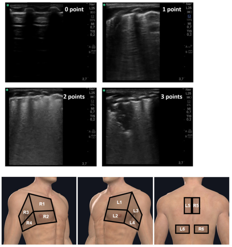
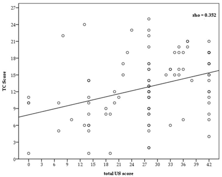
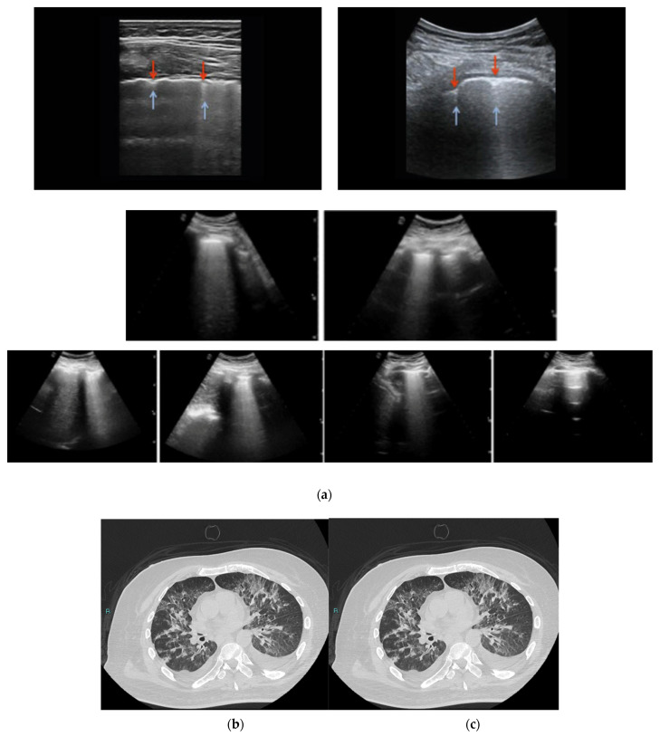
Purpose: We aimed to assess the role of lung ultrasound (LUS) in the diagnosis and prognosis of SARS-CoV-2 pneumonia, by comparing it with High Resolution Computed Tomography (HRCT). Patients and methods: All consecutive patients with laboratory-confirmed SARS-CoV-2 infection and hospitalized in COVID Centers were enrolled. LUS and HRCT were carried out on all patients by expert operators within 48−72 h of admission. A four-level scoring system computed in 12 regions of the chest was used to categorize the ultrasound imaging, from 0 (absence of visible alterations with ultrasound) to 3 (large consolidation and cobbled pleural line). Likewise, a semi-quantitative scoring system was used for HRCT to estimate pulmonary involvement, from 0 (no involvement) to 5 (>75% involvement for each lobe). The total CT score was the sum of the individual lobar scores and ranged from 0 to 25. LUS scans were evaluated according to a dedicated scoring system. CT scans were assessed for typical findings of COVID-19 pneumonia (bilateral, multi-lobar lung infiltration, posterior peripheral ground glass opacities). Oxygen requirement and mortality were also recorded. Results: Ninety-nine patients were included in the study (male 68.7%, median age 71). 40.4% of patients required a Venturi mask and 25.3% required non-invasive ventilation (C-PAP/Bi-level). The overall mortality rate was 21.2% (median hospitalization 30 days). The median ultrasound thoracic score was 28 (IQR 20−36). For the CT evaluation, the mean score was 12.63 (SD 5.72), with most of the patients having LUS scores of 2 (59.6%). The bivariate correlation analysis displayed statistically significant and high positive correlations between both the CT and composite LUS scores and ventilation, lactates, COVID-19 phenotype, tachycardia, dyspnea, and mortality. Moreover, the most relevant and clinically important inverse proportionality in terms of P/F, i.e., a decrease in P/F levels, was indicative of higher LUS/CT scores. Inverse proportionality P/F levels and LUS and TC scores were evaluated by univariate analysis, with a P/F−TC score correlation coefficient of −0.762, p < 0.001, and a P/F−LUS score correlation coefficient of −0.689, p < 0.001. Conclusions: LUS and HRCT show a synergistic role in the diagnosis and disease severity evaluation of COVID-19.
Keywords: ARDS; SARS-CoV-19; high resolution computed tomography; interstitial pneumonia; lung ultrasound.
Conflict of interest statement
The authors declare that the research was conducted in the absence of any commercial or financial relationships that could be construed as a potential conflict of interest.
Figures
Similar articles
-
The value of lung ultrasound in COVID-19 pneumonia, verified by high resolution computed tomography assessed by artificial intelligence.BMC Infect Dis. 2023 Mar 31;23(1):195. doi: 10.1186/s12879-023-08173-4. BMC Infect Dis. 2023. PMID: 37003997 Free PMC article.
-
Lung ultrasound correlates with radiographic severity and pattern in COVID-19 pneumonia: a preliminary study.Ann Palliat Med. 2021 Jul;10(7):8147-8154. doi: 10.21037/apm-21-1731. Ann Palliat Med. 2021. PMID: 34353099
-
Lung Ultrasound in COVID-19 Pneumonia: Correlations with Chest CT on Hospital admission.Respiration. 2020;99(7):617-624. doi: 10.1159/000509223. Epub 2020 Jun 22. Respiration. 2020. PMID: 32570265 Free PMC article.
-
Diagnostic Imaging in Newborns, Children and Adolescents Infected with Severe Acute Respiratory Syndrome Coronavirus 2 (SARS-CoV-2): Is There a Realistic Alternative to Lung High-Resolution Computed Tomography (HRCT) and Chest X-Rays? A Systematic Review of the Literature.Ultrasound Med Biol. 2021 Nov;47(11):3034-3040. doi: 10.1016/j.ultrasmedbio.2021.07.015. Epub 2021 Jul 24. Ultrasound Med Biol. 2021. PMID: 34429231 Free PMC article.
-
Lung ultrasound score severity cut-off points in COVID-19 pneumonia. A systematic review and validating cohort.Med Clin (Barc). 2023 Jun 23;160(12):531-539. doi: 10.1016/j.medcli.2023.01.024. Epub 2023 Mar 10. Med Clin (Barc). 2023. PMID: 36990898 Free PMC article.
Cited by
-
B-Lines Lung Ultrasonography Simulation Using Finite Element Method.Diagnostics (Basel). 2022 Nov 10;12(11):2751. doi: 10.3390/diagnostics12112751. Diagnostics (Basel). 2022. PMID: 36359594 Free PMC article.
-
A Modified Corona Score Using Lung Ultrasound to Identify COVID-19 Patients.Diagnostics (Basel). 2023 Dec 26;14(1):51. doi: 10.3390/diagnostics14010051. Diagnostics (Basel). 2023. PMID: 38201360 Free PMC article.
-
Efficacy of low-intensity pulsed ultrasound in the treatment of COVID-19 pneumonia.Ultraschall Med. 2023 Dec;44(6):e274-e283. doi: 10.1055/a-2133-0835. Epub 2023 Jul 19. Ultraschall Med. 2023. PMID: 37467781 Free PMC article. Clinical Trial.
-
Lung Ultrasound Efficacy in Monitoring Post-SARS-CoV-2 Pneumonia and Inflammatory Biomarkers in Pediatric Patients.Medicina (Kaunas). 2024 Aug 11;60(8):1296. doi: 10.3390/medicina60081296. Medicina (Kaunas). 2024. PMID: 39202577 Free PMC article.
-
COVID-19 Lung Ultrasound Scores and Lessons from the Pandemic: A Narrative Review.Diagnostics (Basel). 2023 Jun 5;13(11):1972. doi: 10.3390/diagnostics13111972. Diagnostics (Basel). 2023. PMID: 37296825 Free PMC article. Review.
References
-
- WHO Coronavirus (COVID-19) Dashboard. [(accessed on 16 February 2022)]; Available online: https://covid19.who.int/
-
- Valdenassi L., Franzini M., Ricevuti G., Rinaldi L., Galoforo A.C., Tirelli U. Letter to the Editor: Potential mechanisms by which the oxygen-ozone (O2–O3) therapy could contribute to the treatment against the coronavirus COVID-19. Eur. Rev. Med. Pharmacol. Sci. 2020;24:4059–4061. doi: 10.26355/eurrev_202004_20976. - DOI - PubMed
-
- Zhou F., Yu T., Du R., Fan G., Liu Y., Liu Z., Xiang J., Wang Y., Song B., Gu X., et al. Clinical Course and Risk Factors for Mortality of Adult Inpatients with COVID-19 in Wuhan, China: A Retrospective Cohort Study. Lancet. 2020;395:1054–1062. doi: 10.1016/S0140-6736(20)30566-3. - DOI - PMC - PubMed
-
- Kudlinski B., Zgoła D., Stolińska M., Murkos M., Kania J., Nowak P., Noga A., Wojciech M., Zaborniak G., Zembron-Lacny A. Systemic Inflammatory Predictors of In-Hospital Mortality in COVID-19 Patients: A Retrospective Study. Diagnostics. 2022;12:859. doi: 10.3390/diagnostics12040859. - DOI - PMC - PubMed
LinkOut - more resources
Full Text Sources
Research Materials
Miscellaneous
