. 2022 Aug 4;12(8):1885.
doi: 10.3390/diagnostics12081885.

Prevalence and Characteristics of Accessory Mandibular Canals: A Cone-Beam Computed Tomography Study in a European Adult Population

Affiliations

Prevalence and Characteristics of Accessory Mandibular Canals: A Cone-Beam Computed Tomography Study in a European Adult Population

Giuseppe Varvara et al. Diagnostics (Basel). .

Abstract

The purpose of this observational study is to evaluate the prevalence and main characteristics of bifid canals within a European adult population, analyzing cone-beam-computed tomography (CBCT). The population study examined 300 subjects. The CBCTs were performed between 2012 and 2019, using PaX-Zenith3D with a standard protocol of acquisition. The parameters analyzed were the presence and lengths of the bifid mandibular canals. The sample included 49% male and 51% female participants. The mean age of the patients was 47.07 ± 17.7 years. Anatomical variants of the mandibular canal were identified in 28.8% of the sides and 50.3% of the patients. In 7.3% of the subjects, the anatomical variants were present bilaterally. The most frequently encountered bifid canal was Type 3 (40.5%), followed by the Type 1 canal (39.3%), the Type 2 canal (14.5%), and the Type 4 canal (5.9%), 40% on the right side and 60% on the left side. The average length of the bifid canals located on the right side of the mandible was 11.96 ± 5.57 mm, compared to 11.38 ± 4.89 mm for those measured on the left side. The bifid mandibular canal is a common anatomical variation of the mandibular canal. It is fundamental to performing an accurate preoperative evaluation using CBCT analysis to avoid and/or reduce intraoperative and postoperative complications.

Keywords: accessory mandibular foramina; bifid canals; inferior alveolar nerve; mandibular canal variant.


Conflict of interest statement

The authors declare no conflict of interest.

Figures

Figure 1
Figure 1
Diagram of the different classifications [12,13,15,16,17].
Figure 2
Figure 2
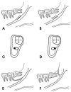
Naitoh classification. The forward canal divided into one with confluence (A) or one without confluence (B). The bucco-lingual canal from the buccal or lingual wall (C,D). The dental canal reaches the root apex (E). The retromolar canal opens into the retromolar region (F). Reprinted with permission from [18] Copyright (2019).
Figure 3
Figure 3
Measuring the length of a Type 1 canal.
Figure 4
Figure 4
Angle measurement of a Type 1 canal.
Figure 5
Figure 5
Type 1, retromolar canal: (A) sagittal view; (B) intraoperative view; (C) Panorex reconstruction; and (D) cross-section reconstruction. Arrow indicated the presence of the canal.
Figure 6
Figure 6
Type 2, dental canal: cross and Panorex reconstructions. Reconstruction shows arrival at the apex of the 4.7 tooth element.
Figure 7
Figure 7
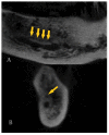
Type 3, confluent anterior canal: (A) Panorex and (B) cross-section reconstructions. Arrows indicate the presence of the canal.
Figure 8
Figure 8
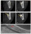
Type 3, not confluent canal: (AD) cross-section reconstructions and (E) Panorex reconstructions. Arrows indicate the presence of the canal.
Figure 9
Figure 9
Type 4: (A) buccal canal cross-section reconstructions and (B) lingual cross-section reconstructions. Arrows indicate the presence of the canal.

References

    1. Manchisi M., Bianchi I., Bernardi S., Varvara G., Pinchi V. Maxillary sinusitis caused by retained dental impression material: An unusual case report and literature review. Niger. J. Clin. Pract. 2022;25:379–385. - PubMed
    1. Carter R.B., Keen E.N. The intramandibular course of the inferior alveolar nerve. J. Anat. 1971;108:433–440. - PMC - PubMed
    1. Bernardi S., Bianchi S., Continenza M.A., Macchiarelli G. Frequency and anatomical features of the mandibular lingual foramina: Systematic review and meta-analysis. Surg. Radiol. Anat. 2017;39:1349–1357. doi: 10.1007/s00276-017-1888-x. - DOI - PubMed
    1. Bogdán S., Pataky L., Barabás J., Németh Z., Huszár T., Szabó G. Atypical courses of the mandibular canal: Comparative examination of dry mandibles and X-rays. J. Craniofac. Surg. 2006;17:487–491. doi: 10.1097/00001665-200605000-00017. - DOI - PubMed
    1. Kuczynski A., Kucharski W., Franco A., Westphalen F.H., de Lima A.A.S., Fernandes A. Prevalence of bifid mandibular canals in panoramic radiographs: A maxillofacial surgical scope. Surg. Radiol. Anat. 2014;36:847–850. doi: 10.1007/s00276-014-1298-2. - DOI - PubMed

LinkOut - more resources