. 2022 Aug 4;12(8):1888.
doi: 10.3390/diagnostics12081888.

MRI Evolution of a Patient with Viral Tick-Borne Encephalitis and Polymorphic Seizures

Affiliations

MRI Evolution of a Patient with Viral Tick-Borne Encephalitis and Polymorphic Seizures

Carmen Adella Sirbu et al. Diagnostics (Basel). .

Abstract

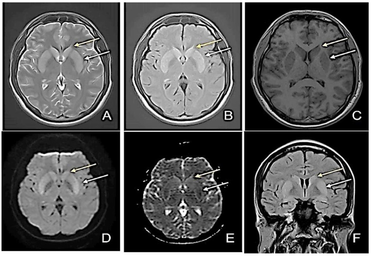
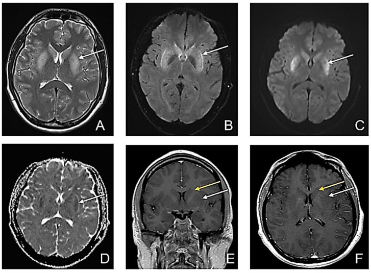
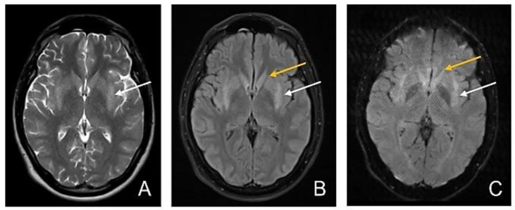
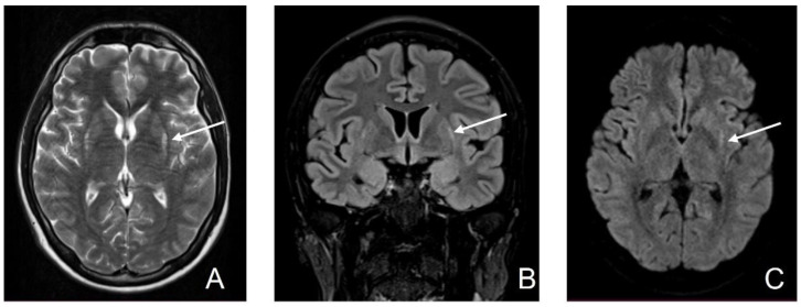
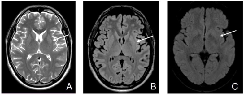
Some neurotropic viruses induce specific lesions in the deep structures, such as basal ganglia and thalamus. These anatomical structures play an important role in initiating and maintaining different types of epileptic seizures. We present the case of a 25-year-old male, transferred to our clinic one week after the onset of the symptomatology, with a recent history of traveling to Turkey and Egypt. At the moment of his hospital admission, his symptoms included altered consciousness, agitation, and seizures. Shortly after, his state worsened, requiring intubation. Viral tick-borne encephalitis diagnoses were favored by the CSF (cerebrospinal fluid) analysis, EEG (Electroencephalography), MRI (magnetic resonance imaging) images presenting symmetric hyper signal in the basal ganglia, and IgM antibodies for anti-tick-borne encephalitis. These lesions persisted for several weeks, and the patient's seizures were polymorphic, originally generalized onset motor, generalized onset non-motor, and focal myoclonic. The patient achieved his independence, seizures decreasing both in intensity and frequency; the MRI images became almost normal. The reduction in antiepileptic doses was not followed by seizure recurrence.

Keywords: CSF; EEG; MRI; basal ganglia; flavivirus; imaging; polymorphic seizures; tick-borne encephalitis (TBE); tick-borne encephalitis virus (TBEV).


Conflict of interest statement

Sirbu CA has received speaker honoraria, consulting fees, travel fees, and research and educational grants from Bayer, Novartis, Merck, Schering, Ever Pharma, Roche, and Teva. Not applicable to the rest of the authors.

Figures

Figure 1
Figure 1
First Magnetic Resonance Imaging (MRI)—day 1 at the admission to our clinic (D1). (A) Axial T2-weighted image sequence: slightly increased bilateral putaminal and caudate nucleus signal; (B) Axial FLAIR sequence: increased bilateral putaminal and caudate nucleus signal; (C) Axial T1-weighted image sequence: slightly decreased bilateral putaminal and caudate nucleus signal; (D) Diffusion-weighted image sequence (DWI); (E) Apparent diffusion coefficient map (ADC): moderate bilateral putaminal restricted diffusion; (F) Coronal FLAIR image: slightly increased bilateral putaminal (white arrow) and caudate nucleus signal (yellow arrow).
Figure 2
Figure 2
Second MRI-D5. (A) Axial T2-weighted image sequence: bilateral putaminal high signal intensity; (B) Axial FLAIR sequence: bilateral putaminal high signal intensity; (C) Axial DWI: high signal intensity in putaminal regions; (D) Axial ADC map: low ADC in the putaminal regions suggestive of restricted diffusion in both putaminal regions (white arrow); (E) Contrast-enhanced Coronal T1wi; and (F) Contrast-enhanced Axial T1wi: no enhancement in the bilateral caudate nucleus (yellow arrow) and putamen (white arrow) with slightly decreased signal in bilateral putamen.
Figure 3
Figure 3
Third MRI—D11. (A) Axial T2-wi sequence: persistent high T2 signal intensity in posterior 2/3 of the putamen; (B) Axial FLAIR image: Symmetrical hyperintensity of the posterior 2/3 of the putamen (white arrow) and normal signal intensity of the caudate nucleus (yellow arrow); (C) Axial DWI: persistent symmetrical moderate restricted diffusion in the posterior 2/3 of the putamen (white arrow) and no restricted diffusion in the caudate nucleus (yellow arrow).
Figure 4
Figure 4
Fourth MRI (D45). (A) T2-wi sequence: limited hyper signal in bilateral putamen in the posterior external region; (B) Cor FLAIR image: minimal hyperintensity in the periphery of bilateral putamen; (C) Axial DWI: minimal restricted diffusion persistent in the periphery of bilateral putamen (white arrow).
Figure 5
Figure 5
Fiveth MRI (D63). (A) Axial T2-wi; (B) Axial FLAIR: normal signal intensity with only a discrete band of peripheral high signal intensity T2 wi/FLAIR in bilateral putamen; (C) Axial DWI: there is no more restricted diffusion in the lenticular nucleus (white arrow).

Similar articles

Cited by

References

    1. Adelshin R.V., Melnikova O.V., Karan L.S., Andaev E., Balakhonov S.V. Complete Genome Sequences of Four European Subtype Strains of Tick-Borne Encephalitis Virus from Eastern Siberia, Russia. Genome Announc. 2015;3:e00609-15. doi: 10.1128/genomeA.00609-15. - DOI - PMC - PubMed
    1. Bogovic P. Tick-borne encephalitis: A review of epidemiology, clinical characteristics, and management. World J. Clin. Cases. 2015;3:430–441. doi: 10.12998/wjcc.v3.i5.430. - DOI - PMC - PubMed
    1. Ruzek D., Županc T.A., Borde J., Chrdle A., Eyer L., Karganova G., Kholodilov I., Knap N., Kozlovskaya L., Matveev A., et al. Tick-borne encephalitis in Europe and Russia: Review of pathogenesis, clinical features, therapy, and vaccines. Antivir. Res. 2019;164:23–51. doi: 10.1016/j.antiviral.2019.01.014. - DOI - PubMed
    1. Romero J.R., Newland J.G. Viral meningitis and encephalitis: Traditional and emerging viral agents. Semin. Pediatr. Infect. Dis. 2003;14:72–82. doi: 10.1053/spid.2003.127223. - DOI - PubMed
    1. d’Orsi G., Martino T., Lalla A., Claudio M.T.D., Carapelle E., Avolio C. Faciobrachial dystonic seizures expressed as epileptic spasms, followed by focal seizures in anti-LGI1 encephalitis: A video-polygraphic study. Epileptic Disord. 2018;20:525–529. doi: 10.1684/epd.2018.1010. - DOI - PubMed

LinkOut - more resources