Review
. 2022 Aug 16;11(16):2541.
doi: 10.3390/cells11162541.

Skin-Aging Pigmentation: Who Is the Real Enemy?

Affiliations
Review

Skin-Aging Pigmentation: Who Is the Real Enemy?

Jin Cheol Kim et al. Cells. .

Abstract

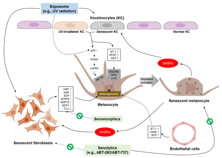
Skin aging is induced and sustained by chronological aging and photoaging. Aging skin pigmentation such as mottled pigmentation (senile lentigo) and melasma are typical signs of photoaging. The skin, like other human organs, undergoes cellular senescence, and senescent cells in the skin increase with age. The crosstalk between melanocytes as pigmentary cells and other adjacent types of aged skin cells such as senescent fibroblasts play a role in skin-aging pigmentation. In this review, we provide an overview of cellular senescence during the skin-aging process. The discussion also includes cellular senescence related to skin-aging pigmentation and the therapeutic potential of regulating the senescence process.

Keywords: aging; cellular senescence; fibroblasts; keratinocytes; melanocytes; senescence; skin pigmentation.


Conflict of interest statement

The authors declare no conflict of interest.

Figures

Figure 1
Figure 1
Cellular crosstalk between melanocytes and neighboring cells in skin-aging pigmentation. UV, ultraviolet; SASP, senescence-associated secretory phenotype, POMC, p53-mediated upregulation of proopiomelanocortin; α-MSH, α-melanocyte-stimulating hormone; MC1R, α-MSH stimulates the melanocortin 1 receptors; ET-1, endothelin-1; bFGF, basic fibroblast growth factor; SCF, stem cell factor; ENDRB, endothelin B receptor; RTK, receptor tyrosinase kinase; HGF, hepatocyte growth factor; KGF, keratinocyte growth factor; sFRP2, secreted frizzled-related protein 2; GDF15, growth differentiation factor 15; SDF1, stromal-derived factor 1; CLU, clusterin; iNOS, inducible nitric oxide synthase.

References

    1. Blokzijl F., de Ligt J., Jager M., Sasselli V., Roerink S., Sasaki N., Huch M., Boymans S., Kuijk E., Prins P., et al. Tissue-specific mutation accumulation in human adult stem cells during life. Nature. 2016;538:260–264. doi: 10.1038/nature19768. - DOI - PMC - PubMed
    1. Herbig U., Ferreira M., Condel L., Carey D., Sedivy J.M. Cellular senescence in aging primates. Science. 2006;311:1257. doi: 10.1126/science.1122446. - DOI - PubMed
    1. Krishnamurthy J., Ramsey M.R., Ligon K.L., Torrice C., Koh A., Bonner-Weir S., Sharpless N.E. p16INK4a induces an age-dependent decline in islet regenerative potential. Nature. 2006;443:453–457. doi: 10.1038/nature05092. - DOI - PubMed
    1. Dimri G.P., Lee X., Basile G., Acosta M., Scott G., Roskelley C., Medrano E.E., Linskens M., Rubelj I., Pereira-Smith O., et al. A biomarker that identifies senescent human cells in culture and in aging skin in vivo. Proc. Natl. Acad. Sci. USA. 1995;92:9363–9367. doi: 10.1073/pnas.92.20.9363. - DOI - PMC - PubMed
    1. Kim T.H., Park T.J., Kweon Y.Y., Baek D.J., Lee J.W., Kang H.Y. Age-Dependent Sequential Increase of Senescent Cells in the Skin. J. Investig. Dermatol. 2022 doi: 10.1016/j.jid.2022.01.027. - DOI - PubMed

Publication types