. 2022 Aug 19;11(16):2584.
doi: 10.3390/cells11162584.

Cellular Therapy Using Epitope-Imprinted Composite Nanoparticles to Remove α-Synuclein from an In Vitro Model

Affiliations

Cellular Therapy Using Epitope-Imprinted Composite Nanoparticles to Remove α-Synuclein from an In Vitro Model

Mei-Hwa Lee et al. Cells. .

Abstract

Several degenerative disorders of the central nervous system, including Parkinson's disease (PD), are related to the pathological aggregation of proteins. Antibodies against toxic disease proteins, such as α-synuclein (SNCA), are therefore being developed as possible therapeutics. In this work, one peptide (YVGSKTKEGVVHGVA) from SNCA was used as the epitope to construct magnetic molecularly imprinted composite nanoparticles (MMIPs). These composite nanoparticles were characterized by dynamic light scattering (DLS), high-performance liquid chromatography (HPLC), isothermal titration calorimetry (ITC), Brunauer-Emmett-Teller (BET) analysis, and superconducting quantum interference device (SQUID) analysis. Finally, the viability of brain endothelial cells that were treated with MMIPs was measured, and the extraction of SNCA from CRISPR/dCas9a-activated HEK293T cells from the in vitro model system was demonstrated for the therapeutic application of MMIPs.

Keywords: gene activation; magnetic nanoparticles; peptide imprinting; protein extraction; α-synuclein.


Conflict of interest statement

The authors declare no conflict of interest.

Figures

Scheme 1
Scheme 1
The preparation of magnetic molecularly imprinted nanoparticles (MMIPs) and their use in the extraction of SNCA from CRISPR/dCas9-activated HEK293T cells.
Figure 1
Figure 1
(a) DLS size distributions of magnetic non- and peptide-imprinted composite nanoparticles before and after peptide removal and rebinding. (b) Mean sizes of nanoparticles prepared with EVALs with various ethylene mol%. AFM images of magnetic (c) non- and (d) peptide-imprinted composite nanoparticles after peptide removal.
Figure 2
Figure 2
(a) BET analysis and specific surface areas (inset) of MNIP and MMIP nanoparticles. (b) SQUID magnetization measurement of MMIPs before and after template removal. MNPs are magnetic nanoparticles alone. (c) Peptide binding to imprinted and non-imprinted nanoparticles, showing increasing imprinting effectiveness with higher ethylene content. (d) Binding isotherm of MNIP and MMIP nanoparticles.
Figure 3
Figure 3
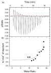
(a) ITC titration curves and (b) the corresponding fitting curve for MMIPs and P5 peptide in water. In this experiment, a suspension of MMIPs (33 μg/mL, 6.6 μM, assuming 0.198 mmol/g binding sites) was titrated with a solution of P5 peptide (1.0 mg/mL: total concentration; 0.66 mM).
Figure 4
Figure 4
(a) Adsorption of P5, P6, and P7 on the P5 MMIPs and MNIPs. (b) Adsorption of P5, P6, and P7 on P5, P6, and P7 MMIPs, respectively. (c) Immunostaining for SNCA in quantum-dot-labeled SNCA recognizing magnetic nanoparticles (QD@MMIP). QD@MMIPs (100 µg/mL) were preincubated with 1, 3, 10, or 30 µg/mL SNCA for 30 min. Scale bar: 10 µm. Images are composites of z-stacks, and thus, the relative intensities are not significant.
Figure 5
Figure 5
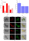
The effect of different concentrations of MMIPs on the (a) cell viability kinetics and (b) cell viability at 24-h time points of endothelial cells. Cultured primary endothelial cells were treated with various concentrations of MMIPs (1 µM to 300 µM) for 24 h. Control group received culture medium. Mean ± SD, n = 4–12, ANOVA, Dunnett’s multiple comparison test, *** p < 0.001 compared to the control group. Cytotoxicity of various concentrations of (c) magnetic CRISPR/Cas9 peptide-imprinted polymers (MQIPs) and (d) magnetic SNCA peptide-imprinted polymers (MMIPs) on HEK293T cells.
Figure 6
Figure 6
(a) Optical, nuclear (DAPI) staining, and immunostaining images of anti-SNCA proteins and merge images of HEK293T cells treated with MNIPs or MMIPs. (b) Optical and immunostaining images of anti-SNCA protein and merge images of MNIPs or MMIPs.

Similar articles

Cited by

References

    1. Chung C.G., Lee H., Lee S.B. Mechanisms of protein toxicity in neurodegenerative diseases. Cell. Mol. Life Sci. 2018;75:3159–3180. doi: 10.1007/s00018-018-2854-4. - DOI - PMC - PubMed
    1. Taylor J.P., Hardy J., Fischbeck K.H. Toxic Proteins in Neurodegenerative Disease. Science. 2002;296:1991–1995. doi: 10.1126/science.1067122. - DOI - PubMed
    1. Carter P.J., Lazar G.A. Next generation antibody drugs: Pursuit of the ‘high-hanging fruit’. Nat. Rev. Drug Discov. 2018;17:197–223. doi: 10.1038/nrd.2017.227. - DOI - PubMed
    1. Sorrentino Z.A., Goodwin M.S., Riffe C.J., Dhillon J.-K.S., Xia Y., Gorion K.-M., Vijayaraghavan N., McFarland K.N., Golbe L.I., Yachnis A.T., et al. Unique α-synuclein pathology within the amygdala in Lewy body dementia: Implications for disease initiation and progression. Acta Neuropathol. Commun. 2019;7:142. doi: 10.1186/s40478-019-0787-2. - DOI - PMC - PubMed
    1. Lang A.E., Lozano A.M. Parkinson’s disease. N. Engl. J. Med. 1998;339:1130–1143. doi: 10.1056/NEJM199810153391607. - DOI - PubMed

Publication types

LinkOut - more resources