Review
. 2022 Aug 20;11(16):2598.
doi: 10.3390/cells11162598.

Glycosylation in Renal Cell Carcinoma: Mechanisms and Clinical Implications

Affiliations
Review

Glycosylation in Renal Cell Carcinoma: Mechanisms and Clinical Implications

Xinqing Zhu et al. Cells. .

Abstract

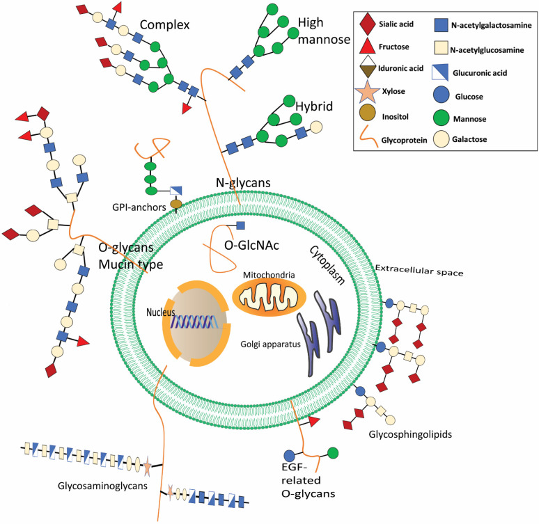
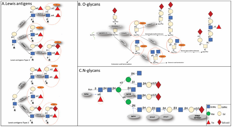
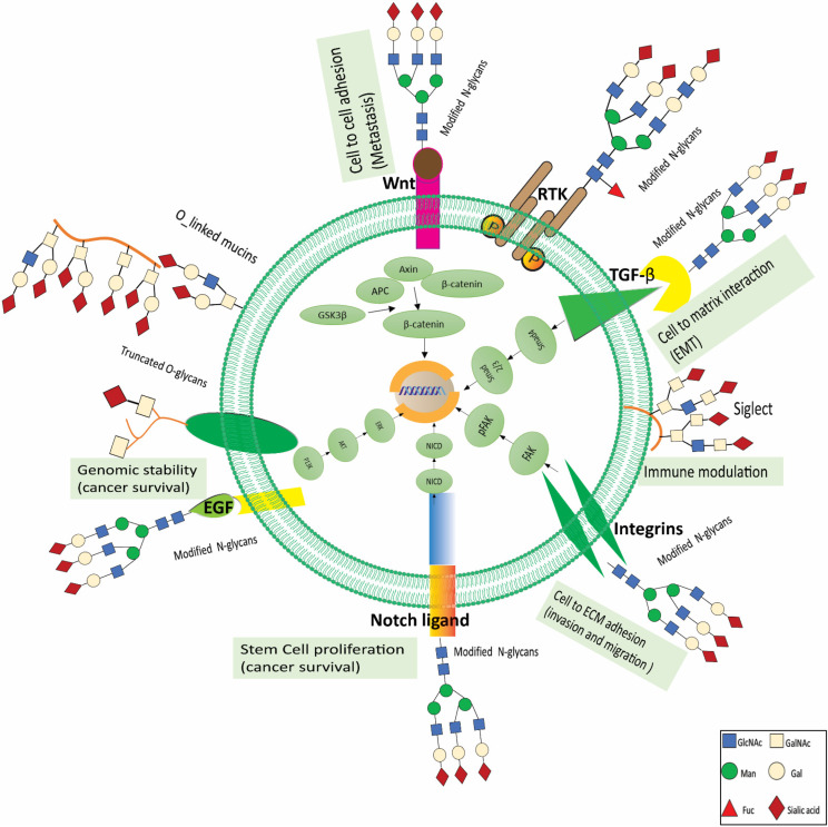
Renal cell carcinoma (RCC) is one of the most prevalent malignant tumors of the urinary system, accounting for around 2% of all cancer diagnoses and deaths worldwide. Clear cell RCC (ccRCC) is the most prevalent and aggressive histology with an unfavorable prognosis and inadequate treatment. Patients' progression-free survival is considerably improved by surgery; however, 30% of patients develop metastases following surgery. Identifying novel targets and molecular markers for RCC prognostic detection is crucial for more accurate clinical diagnosis and therapy. Glycosylation is a critical post-translational modification (PMT) for cancer cell growth, migration, and invasion, involving the transfer of glycosyl moieties to specific amino acid residues in proteins to form glycosidic bonds through the activity of glycosyltransferases. Most cancers, including RCC, undergo glycosylation changes such as branching, sialylation, and fucosylation. In this review, we discuss the latest findings on the significance of aberrant glycans in the initiation, development, and progression of RCC. The potential biomarkers of altered glycans for the diagnosis and their implications in RCC have been further highlighted.

Keywords: biomarkers; fucosylation; glycosylation; glycosyltransferase; renal cell carcinoma; sialylation.


Conflict of interest statement

The authors declare no conflict of interest.

Figures

Figure 1
Figure 1
The most common glycoconjugates present in mammals are depicted. Glycans are present in a wide range of biomolecules. Glycosphingolipids are essential components of the cell’s external membrane. A reversible sequence of structures, called ceramide-linked glycans, are modified by terminal sialic acid. Glycosylation occurs when an N- or O-linked saccharide is attached covalently to Asp or Ser/Thr8 in the polypeptide backbone. N-Glycans all have the same basic structure (N-acetylgalactosamine), but they may be further subdivided into high mannose, hybrid, and complex forms, and their terminal structures can further be modified. When O-linked with Ser/Thr, it will develop mucin-type O, which is often seen in membrane-associated or secreted glycoproteins. Some glycoproteins are also linked to phosphatidylinositol in the plasma membrane’s outer leaflet, called glycosylphosphatidylinositol (GPI) linked proteins. Less common O-glycans may alter epidermal growth factor (EGF)-like repeats (O-fucose, O-glucose, and O-linked N-acetylglucosamine). Glycosaminoglycans are longitudinal co-polymers of acidic disaccharide repeating units found as hyaluronic acid or connected to proteoglycans in the extracellular matrix. Glycosaminoglycans may be sulfated at the N-site (NS) or at multiple O-sites, including 2-O-sulfation (2S), 4-O-sulfation (4S), and 6-O-sulfation (6S). Glycosphingolipids are ceramide-linked glycans that dominate the exterior cellular membranes. In addition, O-GlcNAcylation adds O-linked N-acetylglucosamine to various cytoplasmic and nuclear proteins.
Figure 2
Figure 2
The figure shows some of the important glycan moieties that will be talked about in this study. (A) Terminal Lewis and sialylated Lewis; (B) O-linked and (C) N-linked glycans. Lewis a (Lea), Lewis b (Leb), and sialyl Lewis a (SLea) are type 1 Lewis antigens. Lex, Ley, and SLey are type 2 Lewis antigens. Truncated O-glycans such as T, sialyl Tn(STn), or Tn are often present in cancer cells. Additionally, terminal glycosyl epitopes, such as sialyl Lewis(SLe) epitopes, α 2,3-, or α 2,6-linked sialic acid to N—acetyllactosamine(SLN), and fucosylated Lewis (Le) structures may be found at the ends of the glycan chains. The N-glycan structures of both bisecting N-acetylglucosamine and β 1,6-branching N-acetylglucosamine compete with one another. In cancer, the balance is often tipped in favor of branching. N-Glycans can also be changed by adding fucose to their center. Gangliosides are sialylated glycosphingolipids. Sialylated glycosphingolipids are known as gangliosides. GM3, GD3, or GD2 are often overproduced in the abnormal form in cancer. The key enzymes in charge of adding certain sugar residues are also shown in boxes. GalNAc-T1, GalNAc-T2, GalNAc-T3, GalNAc-T4, GalNAc-T5, and GalNAc-T6 are among the 20 enzymes that make up the family of polypeptide N-acetylgalactosamine transferases (ppGalNA-Ts), sialyltransferases (like-galactoside-2,6 sialyltransferases I (ST6Gal I). FUT8, which helps add “core” 1,6 Fuc to N-glycans; FUT1 and FUT2, that add fucose (Fuc) for 1,2 linkage to galactose (Gal); FUTs that help add Fuc in 1,3 linkage to 2,3 sialylated type 2 chain (FUT3, FUT4, FUT5).
Figure 3
Figure 3
Glycans’ role in RCC growth and progression. Glycans serve critical roles in the pathological stages of tumor formation and progression. Glycans disrupt cell-cell adhesion during cancer cell dissociation and invasion. Epithelial cadherin (E-cadherin) is changed with 1,6-N-acetylglucosamine (1,6GlcNAc)-branched N-glycan structures when the activity of N-acetylglucosaminyltransferase V (GnT V) is elevated. This decreases cell attachment and enhances tumor cell invasion. Such highly branched patterns may be enlarged, and the terminal structures that are 2,6-sialylated inhibit the attachment of tumor cells. Protein stability and the suppression of tumor development may be attributed to the existence of E-cadherin N-glycans that include bisecting GlcNAc structures. This reaction is catalyzed by GnT-III. In addition, abnormal O-glycosylation is associated with tumor cell invasion. One example of this is the production of sialyl Tn (STn), which can be caused by either overexpression of -N-acetylgalactosamine (-GalNAc)-2,6 sialyltransferase I (ST6GalNAc I) or mutations in C1GALTT1-specific chaperone 1 (C1GALT1C1). The formation and proliferation of tumors are characterized by changes in glycosylation of key growth factor receptors, which modifies the receptors’ functions and the signals they send. Ganglioside expression in the membrane of cancer cells may also interfere with signal transmission, setting off a wide array of cellular pathways that foster the growth and development of tumors. Alterations in O-GlcNAcylation have been connected to the development of cancer as well. During the migration of tumor cells, integrins display various glycosylation patterns in O-linked and N-linked glycans. Terminal sialylation disrupts the connections between cells and their extracellular matrix (ECM), which results in an invading and migrating phenotype. The abnormal glycosylation of the vascular endothelial growth factor receptor (VEGFR) changes the way the receptor interacts with galectins and has been associated with the development of tumor angiogenesis. Cancer-associated carbohydrate determinants sialyl lewis x (SLex) and sialyl lewis ea (SLea) function as ligands for adhesion receptors expressed in activated endothelium cells (E-selectin), platelets (P-selectin), and leukocytes (L-selectin), which facilitates cancer cell adherence and metastasis. RTKs are triggered by changes in receptor glycosylation, gangliosides, and glycosaminoglycan expression, resulting in enhanced cancer cell motility, invasion, and proliferation. Fuc stands for fucose; Gal stands for galactose; GlcA stands for glucuronic acid; Man stands for mannose; RTK is for receptor tyrosine kinase; Xyl stands for xylose.

Similar articles

Cited by

References

    1. Siegel R., Naishadham D., Jemal A. Cancer statistics, 2013. CA Cancer J. Clin. 2013;63:11–30. doi: 10.3322/caac.21166. - DOI - PubMed
    1. Siegel R.L., Miller K.D., Jemal A. Cancer statistics, 2020. CA Cancer J. Clin. 2020;70:7–30. doi: 10.3322/caac.21590. - DOI - PubMed
    1. Padala S.A., Barsouk A., Thandra K.C., Saginala K., Mohammed A., Vakiti A., Rawla P., Barsouk A. Epidemiology of Renal Cell Carcinoma. World J. Oncol. 2020;11:79–87. doi: 10.14740/wjon1279. - DOI - PMC - PubMed
    1. Jonasch E., Gao J., Rathmell W.K. Renal cell carcinoma. BMJ (Clin. Res. Ed.) 2014;349:g4797. doi: 10.1136/bmj.g4797. - DOI - PMC - PubMed
    1. Stewart G.D., O’Mahony F.C., Powles T., Riddick A.C., Harrison D.J., Faratian D. What can molecular pathology contribute to the management of renal cell carcinoma? Nat. Rev. Urol. 2011;8:255–265. doi: 10.1038/nrurol.2011.43. - DOI - PubMed